


Function and Biology Details
Reactions catalysed:
L-tyrosine = tyramine + CO(2)
5-hydroxy-L-tryptophan = 5-hydroxytryptamine + CO(2)
Biochemical function:
Biological process:
Cellular component:
Structure analysis Details
Assembly composition:
homo dimer (preferred)
Assembly name:
Tyrosine/DOPA decarboxylase 2 (preferred)
PDBe Complex ID:
PDB-CPX-157101 (preferred)
Entry contents:
1 distinct polypeptide molecule
Macromolecule:
Tyrosine/DOPA decarboxylase 2
Molecule details ›
Chains: A, B, C, D, E, F
Length: 531 amino acids
Theoretical weight: 59.42 KDa
Source organism: Papaver somniferum
Expression system: Escherichia coli
UniProt:
Sequence domains: Pyridoxal-dependent decarboxylase conserved domain
Length: 531 amino acids
Theoretical weight: 59.42 KDa
Source organism: Papaver somniferum
Expression system: Escherichia coli
UniProt:
- Canonical:
 P54769 (Residues: 1-531; Coverage: 100%)
Sequence domains: Pyridoxal-dependent decarboxylase conserved domain
Ligands and Environments
No bound ligands
No modified residues
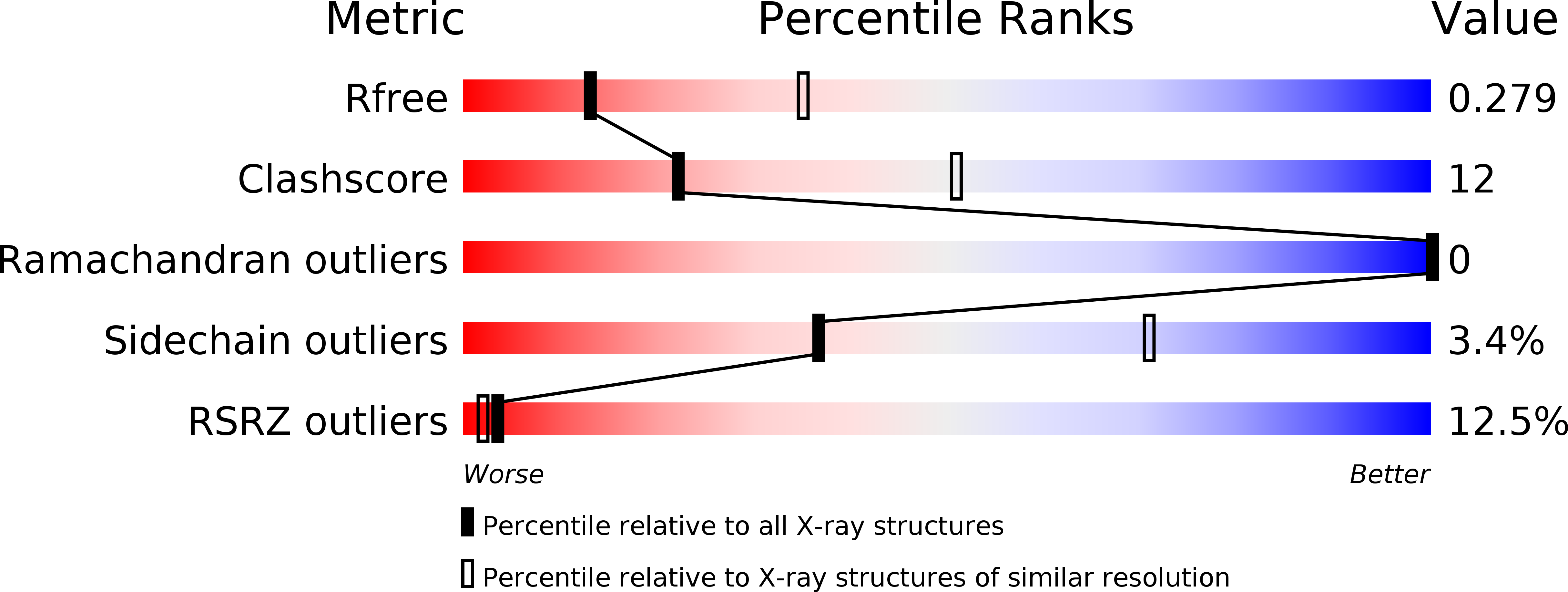
Experiments and Validation Details

X-ray source:
SPRING-8 BEAMLINE BL44XU
Expression system: Escherichia coli