6uuq

X-ray diffraction
1.85Å resolution

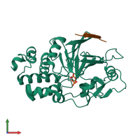
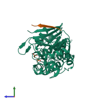
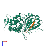
Structure of Calcineurin bound to RCAN1

Released:

Function and Biology Details

Reaction catalysed:
[a protein]-serine/threonine phosphate + H(2)O = [a protein]-serine/threonine + phosphate
Biological process:
Cellular component:
  • not assigned

Structure analysis Details

Assembly composition:
hetero dimer (preferred)
PDBe Complex ID:
PDB-CPX-156877 (preferred)
Entry contents:
2 distinct polypeptide molecules
Macromolecules (2 distinct):
Protein phosphatase 3 catalytic subunit alpha Chain: A
Molecule details ›
Chain: A
Length: 316 amino acids
Theoretical weight: 36.47 KDa
Source organism: Homo sapiens
Expression system: Escherichia coli
UniProt:
  • Canonical: Q08209 (Residues: 26-339; Coverage: 60%)
Gene names: CALNA, CNA, PPP3CA
Sequence domains: Calcineurin-like phosphoesterase
Calcipressin-1 Chain: B
Molecule details ›
Chain: B
Length: 40 amino acids
Theoretical weight: 4.34 KDa
Source organism: Homo sapiens
Expression system: Escherichia coli
UniProt:
  • Canonical: P53805 (Residues: 183-219; Coverage: 15%)
Gene names: ADAPT78, CSP1, DSC1, DSCR1, RCAN1

Ligands and Environments

3 bound ligands:
No modified residues

Experiments and Validation Details

Entry percentile scores
X-ray source: APS BEAMLINE 23-ID-D
Spacegroup: P212121
Unit cell:
a: 57.841Å b: 71.159Å c: 92.049Å
α: 90° β: 90° γ: 90°
R-values:
R R work R free
0.166 0.164 0.205
Expression system: Escherichia coli