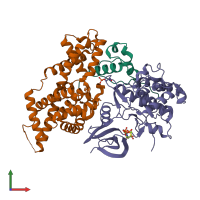
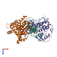
6xbz

Electron Microscopy
2.8Å resolution

Structure of the human CDK-activating kinase

Released:
Source organism: Homo sapiens
Primary publication:
The cryoelectron microscopy structure of the human CDK-activating kinase.
Proc Natl Acad Sci U S A 117 22849-22857 (2020)
PMID: 32855301
Related structures: EMD-22123

Function and Biology Details

Structure analysis Details

Assembly composition:
hetero trimer (preferred)
PDBe Complex ID:
PDB-CPX-156240 (preferred)
Entry contents:
3 distinct polypeptide molecules
Macromolecules (3 distinct):
CDK-activating kinase assembly factor MAT1 Chain: H
Molecule details ›
Chain: H
Length: 328 amino acids
Theoretical weight: 38.13 KDa
Source organism: Homo sapiens
Expression system: Trichoplusia ni
UniProt:
  • Canonical: P51948 (Residues: 1-309; Coverage: 100%)
Gene names: CAP35, MAT1, MNAT1, RNF66
Sequence domains:
Cyclin-H Chain: I
Molecule details ›
Chain: I
Length: 323 amino acids
Theoretical weight: 37.7 KDa
Source organism: Homo sapiens
Expression system: Trichoplusia ni
UniProt:
  • Canonical: P51946 (Residues: 1-323; Coverage: 100%)
Gene name: CCNH
Sequence domains:
Cyclin-dependent kinase 7 Chain: J
Molecule details ›
Chain: J
Length: 391 amino acids
Theoretical weight: 43.73 KDa
Source organism: Homo sapiens
Expression system: Trichoplusia ni
UniProt:
  • Canonical: P50613 (Residues: 1-346; Coverage: 100%)
Gene names: CAK, CAK1, CDK7, CDKN7, MO15, STK1
Sequence domains: Protein kinase domain

Ligands and Environments

2 bound ligands:
1 modified residue:

Experiments and Validation Details

Entry percentile scores
Resolution: 2.8Å
Relevant EMDB volumes: EMD-22123
Expression system: Trichoplusia ni