EMD-1122
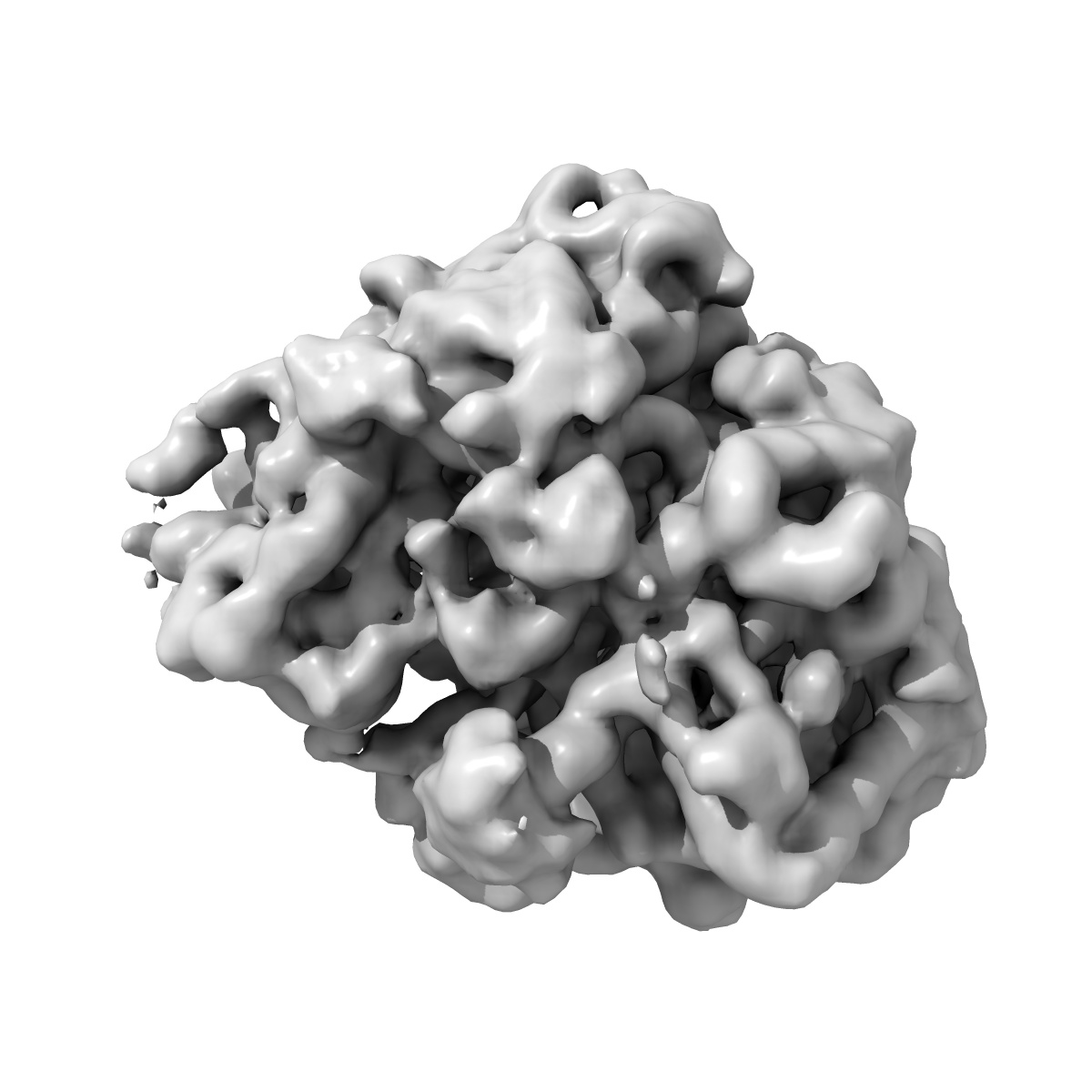
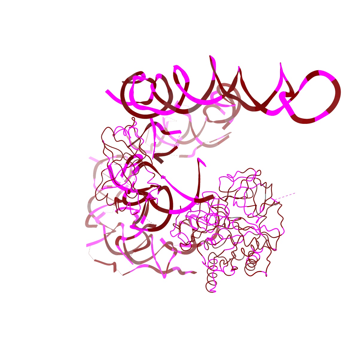
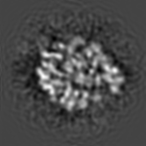
Visualizing tmRNA entry into a stalled ribosome.
EMD-1122
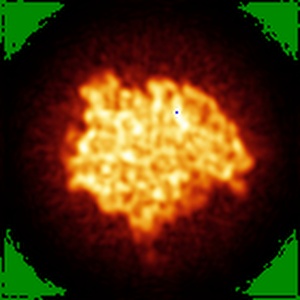
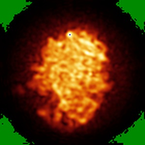
Single-particle13.0 Å
 Deposition: 24/03/2005
Map released: 11/04/2005
Last modified: 24/10/2012
Buffer
pH: 7.5
Details: 25 mM Hepes-KOH pH 7.5, 30 mM NH4Cl, 7mM MgCl2, 2 mM ATP, 6 mM PEP, 10g/ml PK
Details: 25 mM Hepes-KOH pH 7.5, 30 mM NH4Cl, 7mM MgCl2, 2 mM ATP, 6 mM PEP, 10g/ml PK
Vitrification
Cryogen name: ETHANE
Chamber humidity: 90%
Chamber temperature: 93 K
Instrument: HOMEMADE PLUNGER
Method: Blot for 2 seconds before plunging
Details: Vitrification instrument: two side blotting plunger
Chamber humidity: 90%
Chamber temperature: 93 K
Instrument: HOMEMADE PLUNGER
Method: Blot for 2 seconds before plunging
Details: Vitrification instrument: two side blotting plunger
Microscope: FEI TECNAI F20
Illumination mode: FLOOD BEAM
Imaging mode: BRIGHT FIELD
Electron source: FIELD EMISSION GUN
Acceleration voltage: 200 kV
Nominal CS: 2.0 mm
Nominal defocus: 1.45 µm - 3.7 µm
Nominal magnification: 49000.0
Calibrated magnification: 52000.0
Specimen holder model: GATAN LIQUID NITROGEN
Specimen holder details: Cryo transfer
Temperature
Average: 93
K
Image Recording
[1]
Detector category:
FILM
Detector model: KODAK SO-163 FILM
Scanner: ZEISS SCAI
Sampling interval: 14 µm
Number of real images: 44
Average electron dose per image: 15 e/Å2
Old range: 1.2
Bits per pixel: 12.0
Detector model: KODAK SO-163 FILM
Scanner: ZEISS SCAI
Sampling interval: 14 µm
Number of real images: 44
Average electron dose per image: 15 e/Å2
Old range: 1.2
Bits per pixel: 12.0
Format: CCP4
Data type: IMAGE STORED AS FLOATING POINT NUMBER (4 BYTES)
Annotation details: This is a ribosome consisting 70S + Psite tRNA + mRNA + EF-Tu.GDP + alanylated tmRNA + SmpB + Kirromycin + S1
Details: ::::EMDATABANK.org::::EMD-1122::::
Data type: IMAGE STORED AS FLOATING POINT NUMBER (4 BYTES)
Annotation details: This is a ribosome consisting 70S + Psite tRNA + mRNA + EF-Tu.GDP + alanylated tmRNA + SmpB + Kirromycin + S1
Details: ::::EMDATABANK.org::::EMD-1122::::
⬡ Geometry
| X | Y | Z | |
|---|---|---|---|
| Dimensions | 140 | 140 | 140 |
| Origin | -70 | -70 | -70 |
| Spacing | 140 | 140 | 140 |
| Voxel size | 2.82 Å | 2.82 Å | 2.82 Å |
Contour list
| Primary | Level | Source |
|---|---|---|
| True | 54.9 | - |