EMD-1615
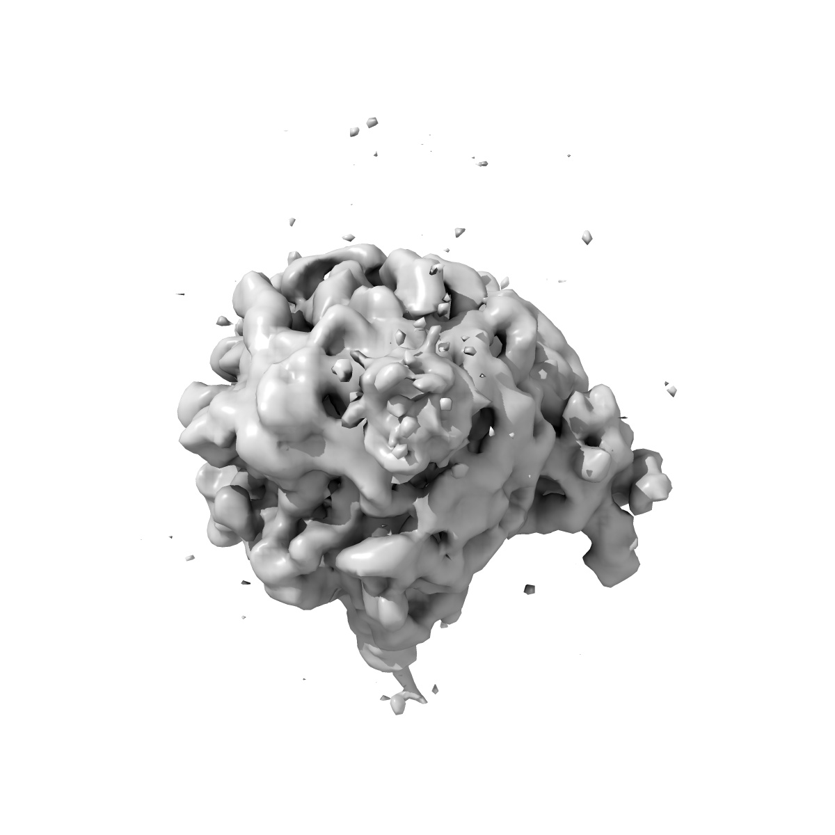
Three-dimensional structure of YidC bound to the translating ribosome
EMD-1615
Single-particle14.4 Å
 Deposition: 23/04/2009
Map released: 10/09/2010
Last modified: 11/12/2013
Grid
Details: 200 mesh copper
Vitrification
Microscope: FEI TECNAI 20
Illumination mode: FLOOD BEAM
Imaging mode: BRIGHT FIELD
Electron source: FIELD EMISSION GUN
Acceleration voltage: 200 kV
Nominal defocus: 1.0 µm - 4.0 µm
Nominal magnification: 50000.0
Calibrated magnification: 50000.0
Specimen holder model: GATAN LIQUID NITROGEN
Specimen holder details: Eucentric
Minimum tilt angle: 0
Maximum tilt angle: 0
Image Recording
[1]
Detector category:
FILM
Detector model: KODAK SO-163 FILM
Scanner: NIKON SUPER COOLSCAN 9000
Sampling interval: 12.7 µm
Average electron dose per image: 15 e/Å2
Bits per pixel: 16.0
Detector model: KODAK SO-163 FILM
Scanner: NIKON SUPER COOLSCAN 9000
Sampling interval: 12.7 µm
Average electron dose per image: 15 e/Å2
Bits per pixel: 16.0
Final
reconstruction
Resolution: 14.4
Å
(
BY AUTHOR)
Resolution method: OTHER
Number of images used: 24395
Algorithm: OTHER
Resolution method: OTHER
Number of images used: 24395
Algorithm: OTHER
⌯ Applied Symmetry
Point group:
C1
Software
[1]
| Name | Version | Details |
|---|---|---|
| Imagic-5 Spider | - | - |
CTF correction
Details:Each image
Format: CCP4
Data type: IMAGE STORED AS FLOATING POINT NUMBER (4 BYTES)
Annotation details: This map shows YidC bound to the tunnel exit region of an ribosome nascent chain complex
Details: ::::EMDATABANK.org::::EMD-1615::::
Data type: IMAGE STORED AS FLOATING POINT NUMBER (4 BYTES)
Annotation details: This map shows YidC bound to the tunnel exit region of an ribosome nascent chain complex
Details: ::::EMDATABANK.org::::EMD-1615::::
⬡ Geometry
| X | Y | Z | |
|---|---|---|---|
| Dimensions | 108 | 108 | 108 |
| Origin | 0 | 0 | 0 |
| Spacing | 108 | 108 | 108 |
| Voxel size | 3.76 Å | 3.76 Å | 3.76 Å |
Contour list
| Primary | Level | Source |
|---|---|---|
| True | 40.0 | AUTHOR |