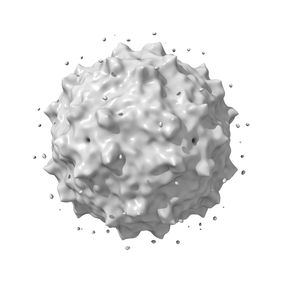
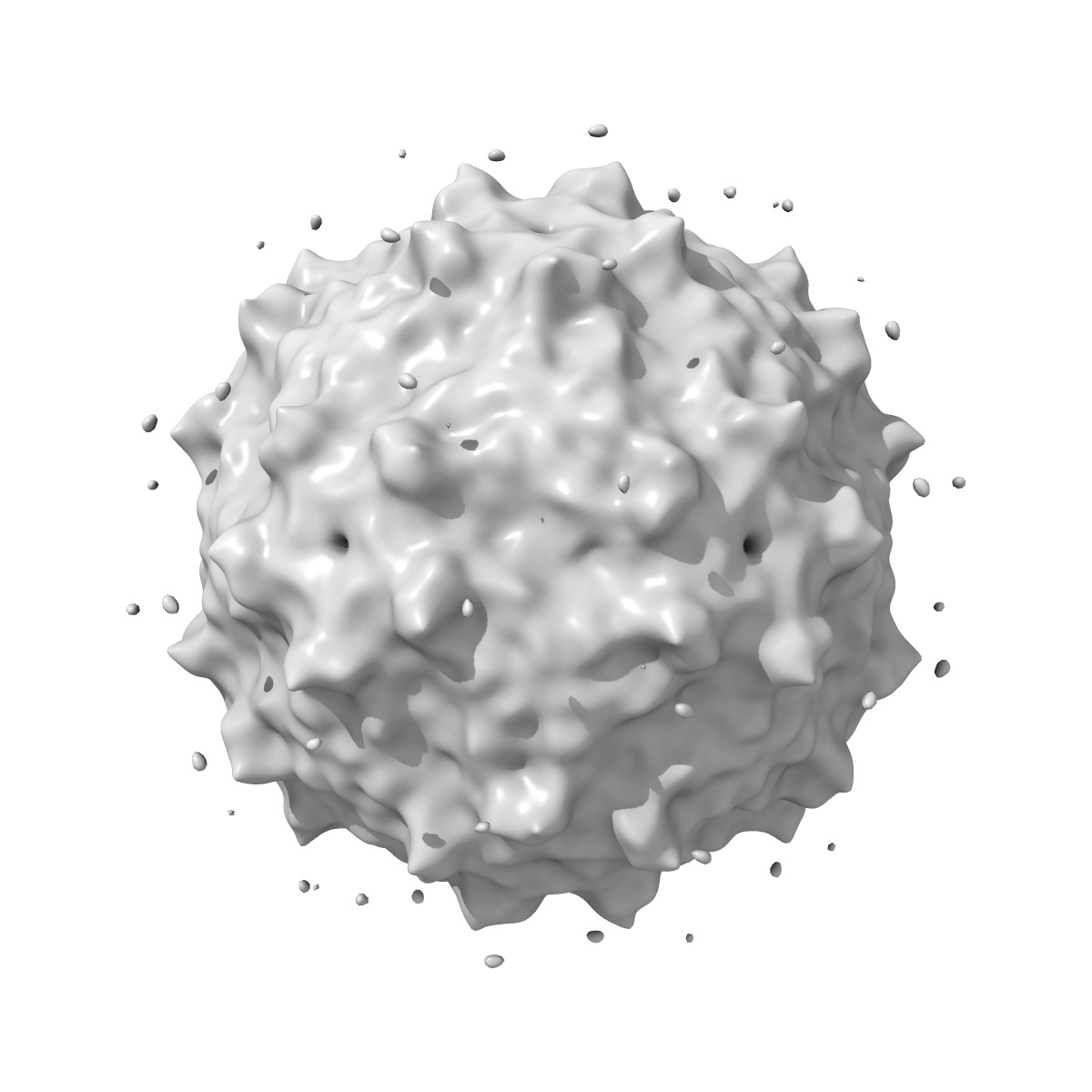
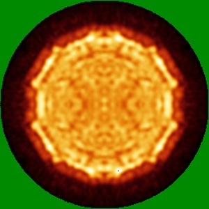
EMD-2025
CryoEM 3D reconstruction of full Triatoma Virus
EMD-2025
Single-particle15.0 Å
 Deposition: 06/01/2012
Map released: 23/01/2013
Last modified: 08/10/2014
Microscope: JEOL 2010F
Illumination mode: OTHER
Imaging mode: BRIGHT FIELD
Electron source: FIELD EMISSION GUN
Acceleration voltage: 200 kV
Nominal CS: 1.4 mm
Nominal defocus: 1.2 µm - 4.0 µm
Nominal magnification: 40000.0
Specimen holder model: GATAN LIQUID NITROGEN
Specimen holder details: Side entry liquid nitrogen-cooled cryo specimen holder
Image Recording
[1]
Detector category:
FILM
Detector model: KODAK SO-163 FILM
Scanner: ZEISS SCAI
Sampling interval: 7 µm
Number of real images: 25
Detector model: KODAK SO-163 FILM
Scanner: ZEISS SCAI
Sampling interval: 7 µm
Number of real images: 25
Final
reconstruction
Resolution: 15.0
Å
(
BY AUTHOR)
Resolution method: FSC 0.5 CUT-OFF
Number of images used: 1716
Resolution method: FSC 0.5 CUT-OFF
Number of images used: 1716
⌯ Applied Symmetry
Point group:
I
Software
[1]
| Name | Version | Details |
|---|---|---|
| EM3DR2 | - | - |
Format: CCP4
Data type: IMAGE STORED AS FLOATING POINT NUMBER (4 BYTES)
Annotation details: Full TrV particle 3D reconstruction
Details: ::::EMDATABANK.org::::EMD-2025::::
Data type: IMAGE STORED AS FLOATING POINT NUMBER (4 BYTES)
Annotation details: Full TrV particle 3D reconstruction
Details: ::::EMDATABANK.org::::EMD-2025::::
⬡ Geometry
| X | Y | Z | |
|---|---|---|---|
| Dimensions | 255 | 255 | 255 |
| Origin | -127 | -127 | -127 |
| Spacing | 255 | 255 | 255 |
| Voxel size | 1.75 Å | 1.75 Å | 1.75 Å |
Contour list
| Primary | Level | Source |
|---|---|---|
| True | 37.0 | AUTHOR |