EMD-2159
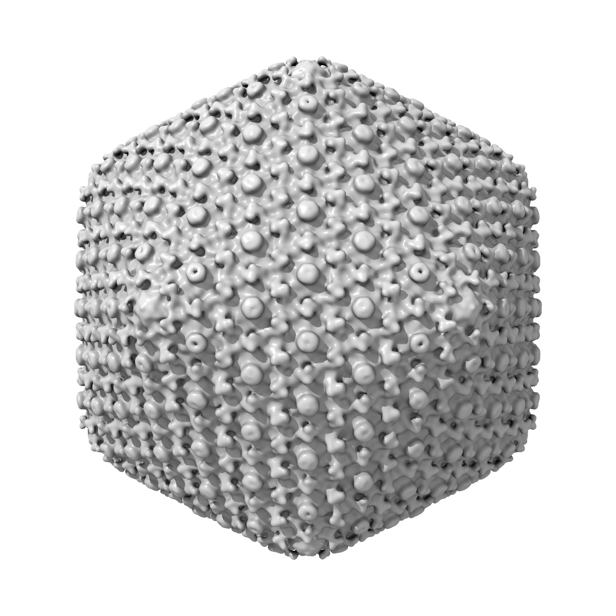
Electron cryo-microscopy of the intact (DNA-filled) head of the yersiniophage phiR1-37
EMD-2159
Single-particle23.8 Å
 Deposition: 09/07/2012
Map released: 03/10/2012
Last modified: 24/10/2012
Buffer
Details: 50 mM Tris pH 7.8, 10 mM MgSO4
Vitrification
Microscope: FEI TECNAI F20
Illumination mode: FLOOD BEAM
Imaging mode: BRIGHT FIELD
Electron source: FIELD EMISSION GUN
Acceleration voltage: 200 kV
Nominal CS: 2.0 mm
Nominal defocus: 0.5 µm - 5.76 µm
Nominal magnification: 62000.0
Specimen holder model: GATAN LIQUID NITROGEN
Specimen holder details: Side entry liquid nitrogen-cooled cryo specimen holder
Details: Low dose conditions
Image Recording
[1]
Detector category:
FILM
Detector model: KODAK SO-163 FILM
Scanner: ZEISS SCAI
Number of real images: 297
Average electron dose per image: 15 e/Å2
Detector model: KODAK SO-163 FILM
Scanner: ZEISS SCAI
Number of real images: 297
Average electron dose per image: 15 e/Å2
Final
reconstruction
Resolution: 23.8
Å
(
BY AUTHOR)
Resolution method: FSC 0.5 CUT-OFF
Number of images used: 1028
Resolution method: FSC 0.5 CUT-OFF
Number of images used: 1028
⌯ Applied Symmetry
Point group:
I
Software
[1]
| Name | Version | Details |
|---|---|---|
| AUTO3DEM | - | - |
CTF correction
Details:Each micrograph
Format: CCP4
Data type: IMAGE STORED AS SIGNED INTEGER (2 BYTES)
Annotation details: Three-dimensional reconstruction of the intact (DNA-filled) yersiniophage phiR1-37
Details: ::::EMDATABANK.org::::EMD-2159::::
Data type: IMAGE STORED AS SIGNED INTEGER (2 BYTES)
Annotation details: Three-dimensional reconstruction of the intact (DNA-filled) yersiniophage phiR1-37
Details: ::::EMDATABANK.org::::EMD-2159::::
⬡ Geometry
| X | Y | Z | |
|---|---|---|---|
| Dimensions | 401 | 401 | 401 |
| Origin | 0 | 0 | 0 |
| Spacing | 401 | 401 | 401 |
| Voxel size | 4.516 Å | 4.516 Å | 4.516 Å |
Contour list
| Primary | Level | Source |
|---|---|---|
| True | 12000.0 | AUTHOR |