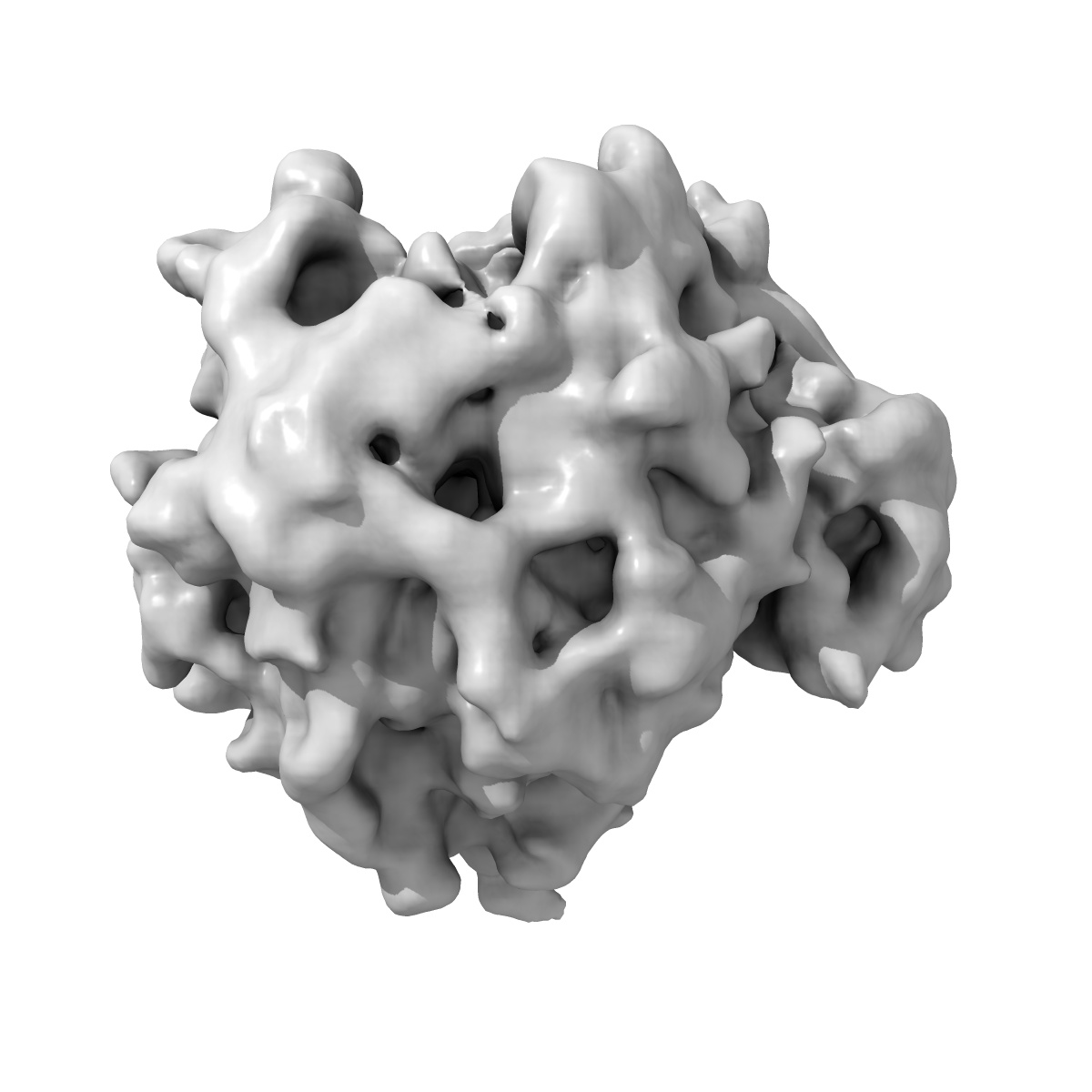
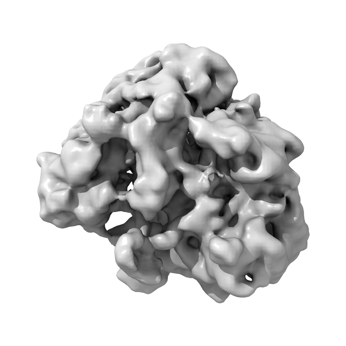
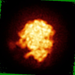
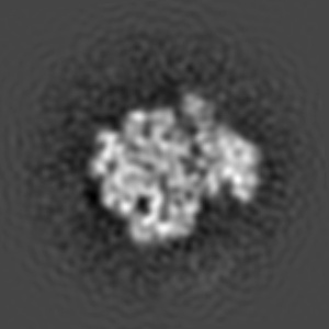
EMD-2172
Methanococcus igneus 70S ribosome
EMD-2172
Single-particle18.0 Å
 Deposition: 09/08/2012
Map released: 13/02/2013
Last modified: 13/02/2013
Buffer
pH: 7.5
Staining
Type:
NEGATIVE
Details: Blot for 10 seconds before plunging, use 2 layers of filter paper
Details: Blot for 10 seconds before plunging, use 2 layers of filter paper
Vitrification
Cryogen name: ETHANE
Chamber humidity: 100%
Instrument: FEI VITROBOT MARK III
Method: Blot for 10 seconds before plunging, use 2 layers of filter paper
Details: Vitrification instrument: Vitrobot
Chamber humidity: 100%
Instrument: FEI VITROBOT MARK III
Method: Blot for 10 seconds before plunging, use 2 layers of filter paper
Details: Vitrification instrument: Vitrobot
Microscope: FEI TECNAI 12
Illumination mode: FLOOD BEAM
Imaging mode: BRIGHT FIELD
Electron source: LAB6
Acceleration voltage: 120 kV
Nominal CS: 2.0 mm
Specimen holder model: GATAN LIQUID NITROGEN
Illumination mode: FLOOD BEAM
Imaging mode: BRIGHT FIELD
Electron source: LAB6
Acceleration voltage: 120 kV
Nominal CS: 2.0 mm
Specimen holder model: GATAN LIQUID NITROGEN
Image Recording
[1]
Detector category:
FILM
Detector model: FEI EAGLE (4k x 4k)
Scanner: OTHER
Number of real images: 99
Average electron dose per image: 20 e/Å2
Details: Data collected on CCD
Detector model: FEI EAGLE (4k x 4k)
Scanner: OTHER
Number of real images: 99
Average electron dose per image: 20 e/Å2
Details: Data collected on CCD
Final
reconstruction
Resolution: 18.0
Å
(
BY AUTHOR)
Resolution method: FSC 0.5 CUT-OFF
Number of images used: 8932
Algorithm: OTHER
Resolution method: FSC 0.5 CUT-OFF
Number of images used: 8932
Algorithm: OTHER
⌯ Applied Symmetry
Point group:
C1
Software
[1]
| Name | Version | Details |
|---|---|---|
| SPIDER | - | - |
CTF correction
Details:Wiener filter on 3D volumes
Format: CCP4
Data type: IMAGE STORED AS FLOATING POINT NUMBER (4 BYTES)
Annotation details: Methanococcus igneus 70S ribosome
Details: ::::EMDATABANK.org::::EMD-2172::::
Data type: IMAGE STORED AS FLOATING POINT NUMBER (4 BYTES)
Annotation details: Methanococcus igneus 70S ribosome
Details: ::::EMDATABANK.org::::EMD-2172::::
⬡ Geometry
| X | Y | Z | |
|---|---|---|---|
| Dimensions | 368 | 368 | 368 |
| Origin | -184 | -184 | -183 |
| Spacing | 368 | 368 | 368 |
| Voxel size | 3.31 Å | 3.31 Å | 3.31 Å |
Contour list
| Primary | Level | Source |
|---|---|---|
| True | 2.17 | EMDB |