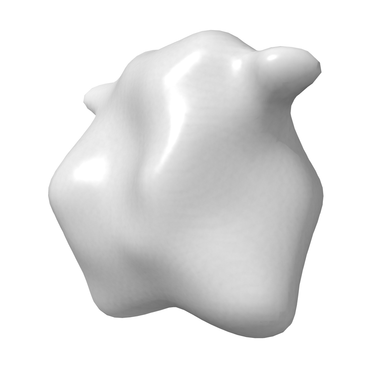
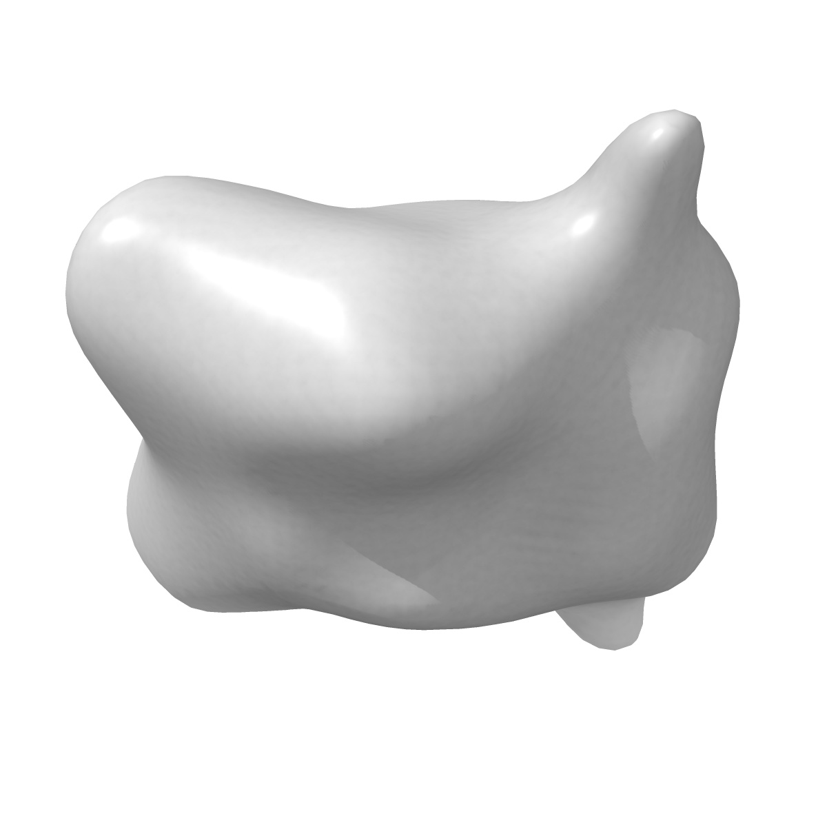
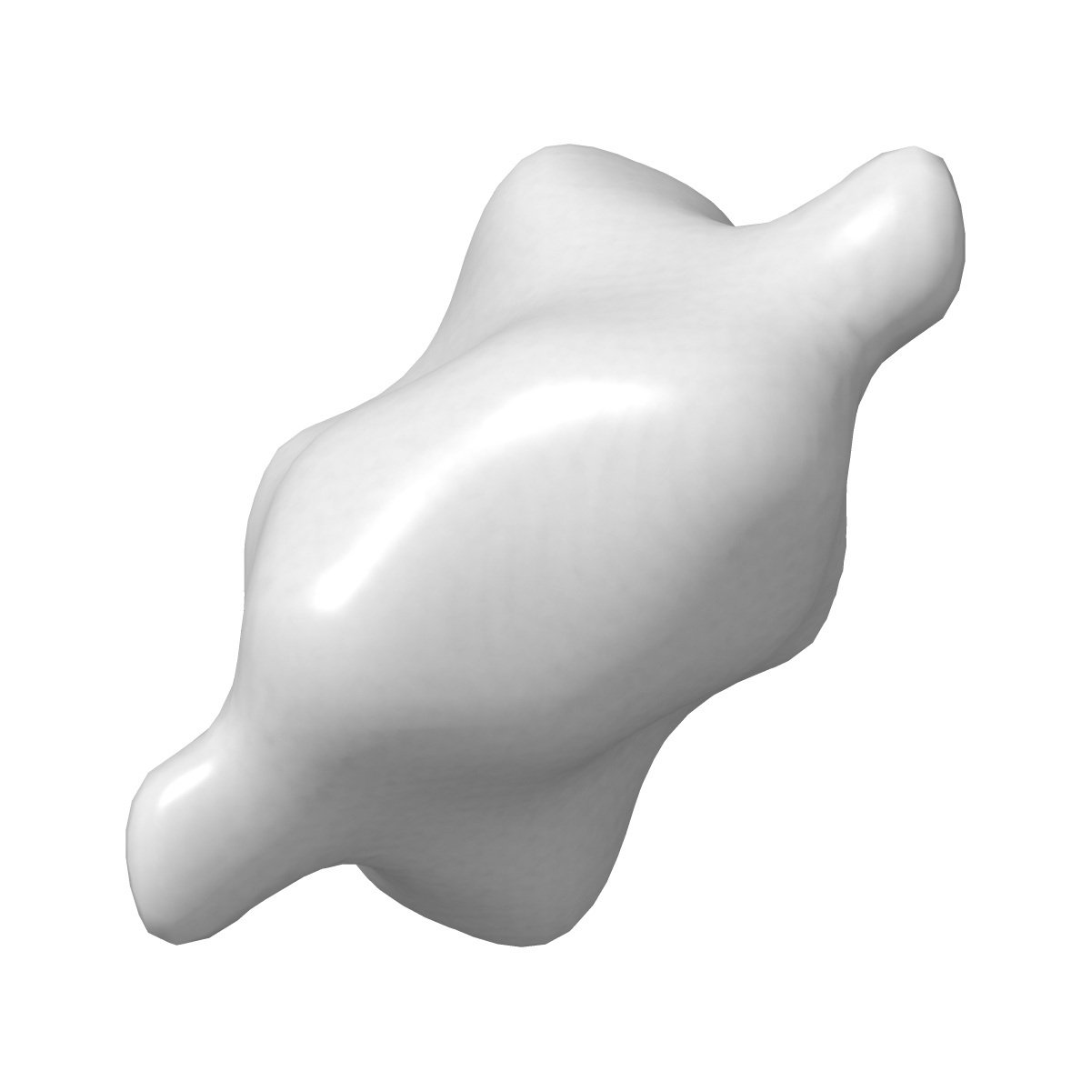
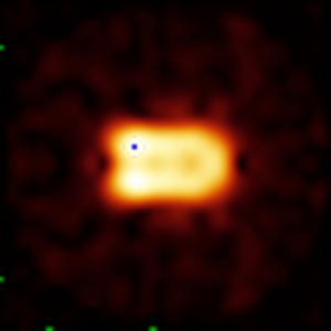
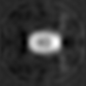
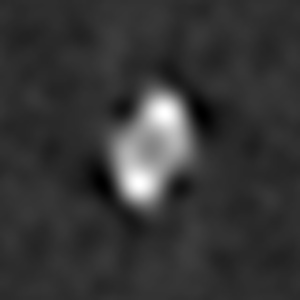
EMD-2182
Electron microscopy map of SOS1 antiporter
EMD-2182
Single-particle25.9 Å
 Deposition: 24/08/2012
Map released: 03/10/2012
Last modified: 28/11/2012
Buffer
pH: 8.5
Details: 50 mM phosphate buffer pH 8.5, 300 mM NaCl, 10% glycerol, 0.05% DDM, 250 mM imidazole
Details: 50 mM phosphate buffer pH 8.5, 300 mM NaCl, 10% glycerol, 0.05% DDM, 250 mM imidazole
Staining
Type:
NEGATIVE
Details: Purified SOS1 was adsorbed to glow-discharged carbon coated grids and stained with 2% uranyl formate
Details: Purified SOS1 was adsorbed to glow-discharged carbon coated grids and stained with 2% uranyl formate
Grid
Details: 400 mesh carbon coated copper grid, glow discharged in moderate vacuum
Microscope: JEOL 2100
Illumination mode: FLOOD BEAM
Imaging mode: BRIGHT FIELD
Electron source: LAB6
Acceleration voltage: 200 kV
Nominal CS: 1.0 mm
Nominal defocus: 1.0 µm - 2.5 µm
Nominal magnification: 20000.0
Calibrated magnification: 20000.0
Specimen holder model: JEOL
Alignment procedure: LEGACY (Astigmatism: Objective lens astigmatism was corrected at 120.000-150.000 times magnification and confirmed by inspection of the Fourier transform, Electron beam tilt params: )
Minimum tilt angle: 0
Maximum tilt angle: 0
Image Recording
[1]
Detector category:
CCD
Detector model: GATAN ORIUS SC200 (2k x 2k)
Number of real images: 90
Average electron dose per image: 15 e/Å2
Bits per pixel: 14.0
Detector model: GATAN ORIUS SC200 (2k x 2k)
Number of real images: 90
Average electron dose per image: 15 e/Å2
Bits per pixel: 14.0
Details: Single images of SOS1 were manually extracted using EMAN. 2D reference-free classification, 2D averaging, 3D classification and reconstruction were performed using maximum-likelihood methods implemented in XMIPP package.
Final
reconstruction
Resolution: 25.9
Å
(
BY AUTHOR)
Resolution method: FSC 0.5 CUT-OFF
Number of images used: 6300
Algorithm: OTHER
Details:
Resolution method: FSC 0.5 CUT-OFF
Number of images used: 6300
Algorithm: OTHER
Details:
⌯ Applied Symmetry
Point group:
C2
Software
[1]
| Name | Version | Details |
|---|---|---|
| XMIPP | - | - |
Final 2D classification
Number of classes:
96
CTF correction
Details:Each micrograph. Estimation using CTFFIND3 and correction using Bsoft
Format: CCP4
Data type: IMAGE STORED AS FLOATING POINT NUMBER (4 BYTES)
Annotation details: SOS1 antiporter class 2
Details: ::::EMDATABANK.org::::EMD-2182::::
Data type: IMAGE STORED AS FLOATING POINT NUMBER (4 BYTES)
Annotation details: SOS1 antiporter class 2
Details: ::::EMDATABANK.org::::EMD-2182::::
⬡ Geometry
| X | Y | Z | |
|---|---|---|---|
| Dimensions | 80 | 80 | 80 |
| Origin | 1 | 1 | 1 |
| Spacing | 80 | 80 | 80 |
| Voxel size | 3.3 Å | 3.3 Å | 3.3 Å |
Contour list
| Primary | Level | Source |
|---|---|---|
| True | 0.035 | AUTHOR |