EMD-2195
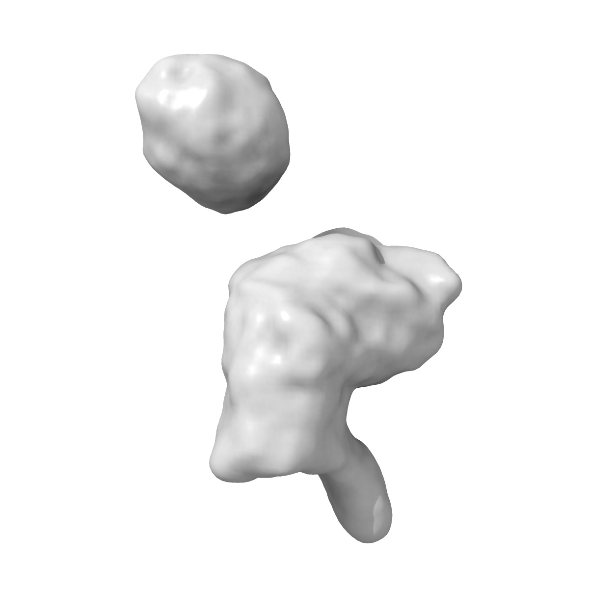
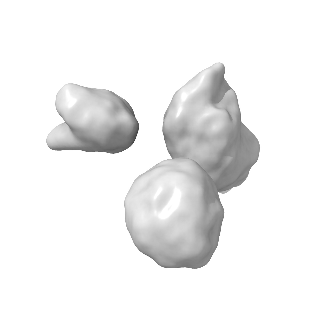
Characterization of the insertase for beta-barrel proteins of the outer mitochondrial membrane. 3-D reconstruction of the TOB complex
EMD-2195
Single-particle14.7 Å
 Deposition: 11/09/2012
Map released: 14/11/2012
Last modified: 17/07/2013
Method: Single-particle
Concentration: 1
mg/mL
Buffer
pH: 8.5
Details: 1mM PMSF, 0.08%(v/v) Triton X-100, 50 mM HEPES pH 8.5
Details: 1mM PMSF, 0.08%(v/v) Triton X-100, 50 mM HEPES pH 8.5
Grid
Details: Lacey carbon films on 200 mesh Molybdenum grids
Vitrification
Cryogen name: ETHANE
Chamber temperature: 120 K
Instrument: HOMEMADE PLUNGER
Method: blot for 4-5 seconds before plunging with whatman filter paper
Chamber temperature: 120 K
Instrument: HOMEMADE PLUNGER
Method: blot for 4-5 seconds before plunging with whatman filter paper
Microscope: FEI TECNAI F20
Illumination mode: FLOOD BEAM
Imaging mode: BRIGHT FIELD
Electron source: FIELD EMISSION GUN
Acceleration voltage: 120 kV
Nominal CS: 2.0 mm
Nominal defocus: -0.5 µm - -3.7 µm
Nominal magnification: 62000.0
Calibrated magnification: 84270.0
Specimen holder model: GATAN LIQUID NITROGEN
Specimen holder details: Gatan 656 side entry holder
Alignment procedure: LEGACY (Astigmatism: Objective lens astigmatism was corrected using the live FFT at imaging magnification., Electron beam tilt params: )
Minimum tilt angle: 0
Maximum tilt angle: 0
Temperature
Maximum: 95
K
Image Recording
[1]
Detector category:
CCD
Detector model: FEI EAGLE (4k x 4k)
Number of real images: 546
Average electron dose per image: 20 e/Å2
Details: Images collected using TOM_acquisition software. 546 good micrographs were CTF corrected for phase. 175,000 particles were automatically selected. In the end 76700 were included in the final reconstruction.
Final
reconstruction
Resolution: 14.7
Å
(
BY AUTHOR)
Resolution method: FSC 0.5 CUT-OFF
Number of images used: 1
Algorithm: OTHER
Resolution method: FSC 0.5 CUT-OFF
Number of images used: 1
Algorithm: OTHER
⌯ Applied Symmetry
Point group:
C1
Software
[1]
| Name | Version | Details |
|---|---|---|
| SPIDER, TOM_toolbox | - | - |
CTF correction
Details:Phase and astigmatism correction applied to each micrograph
Format: CCP4
Data type: IMAGE STORED AS FLOATING POINT NUMBER (4 BYTES)
Annotation details: 3D reconstruction of TOB complexes isolated using the 9xHis tag on the Tob37 subunit. The TOB complex is a hetero trimer containing one copy each of Tob37, Tob38 and Tob37. Total mol. Wgt. Is 140 kDa. In this sample there are three isoforms of Tob55 54.7, 54.1 and 50.7 kDa respectively. This affects the reconstruction.
Details: ::::EMDATABANK.org::::EMD-2195::::
⬡ Geometry
| X | Y | Z | |
|---|---|---|---|
| Dimensions | 140 | 140 | 140 |
| Origin | 0 | 0 | 0 |
| Spacing | 140 | 140 | 140 |
| Voxel size | 1.78 Å | 1.78 Å | 1.78 Å |
Contour list
| Primary | Level | Source |
|---|---|---|
| True | 0.08 | AUTHOR |