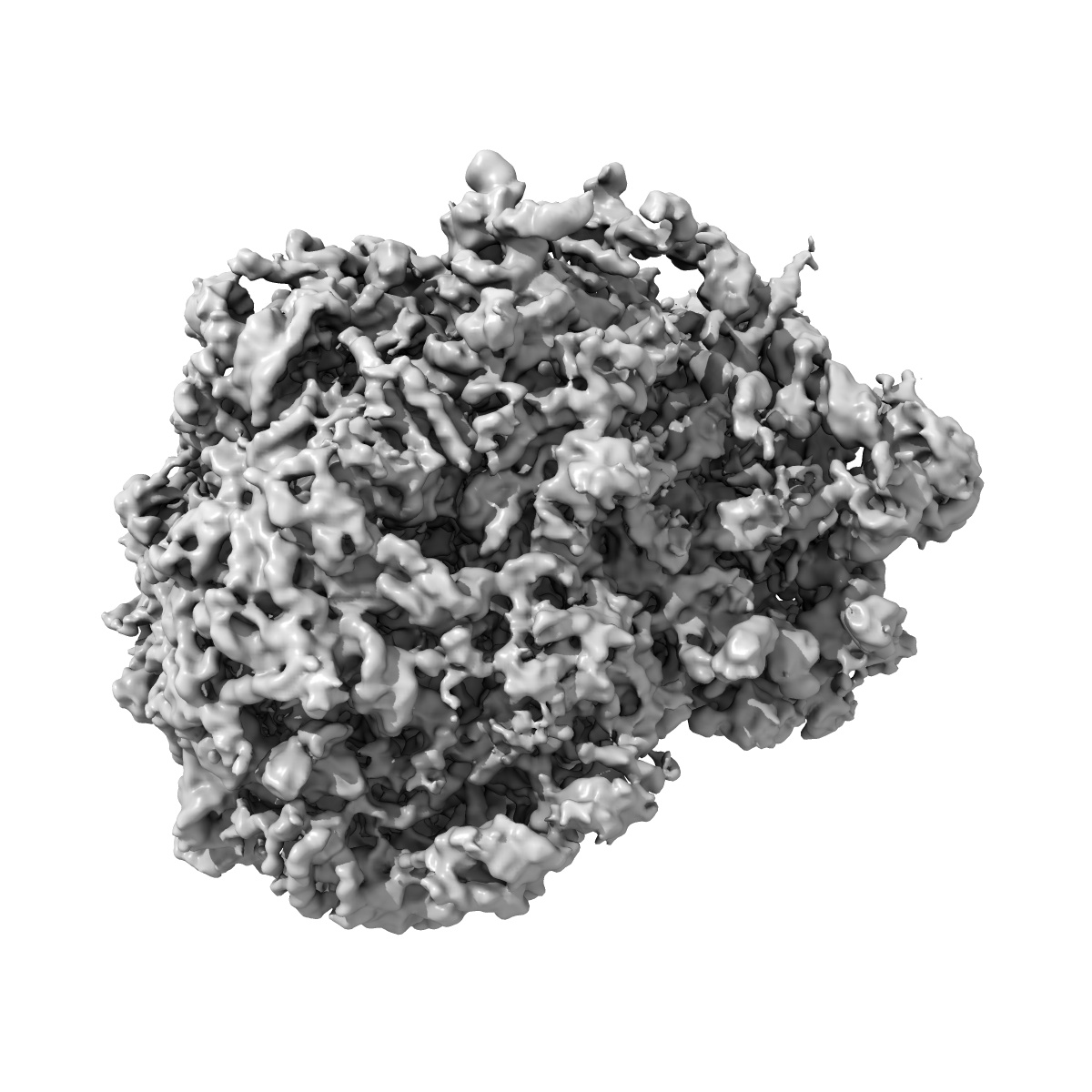
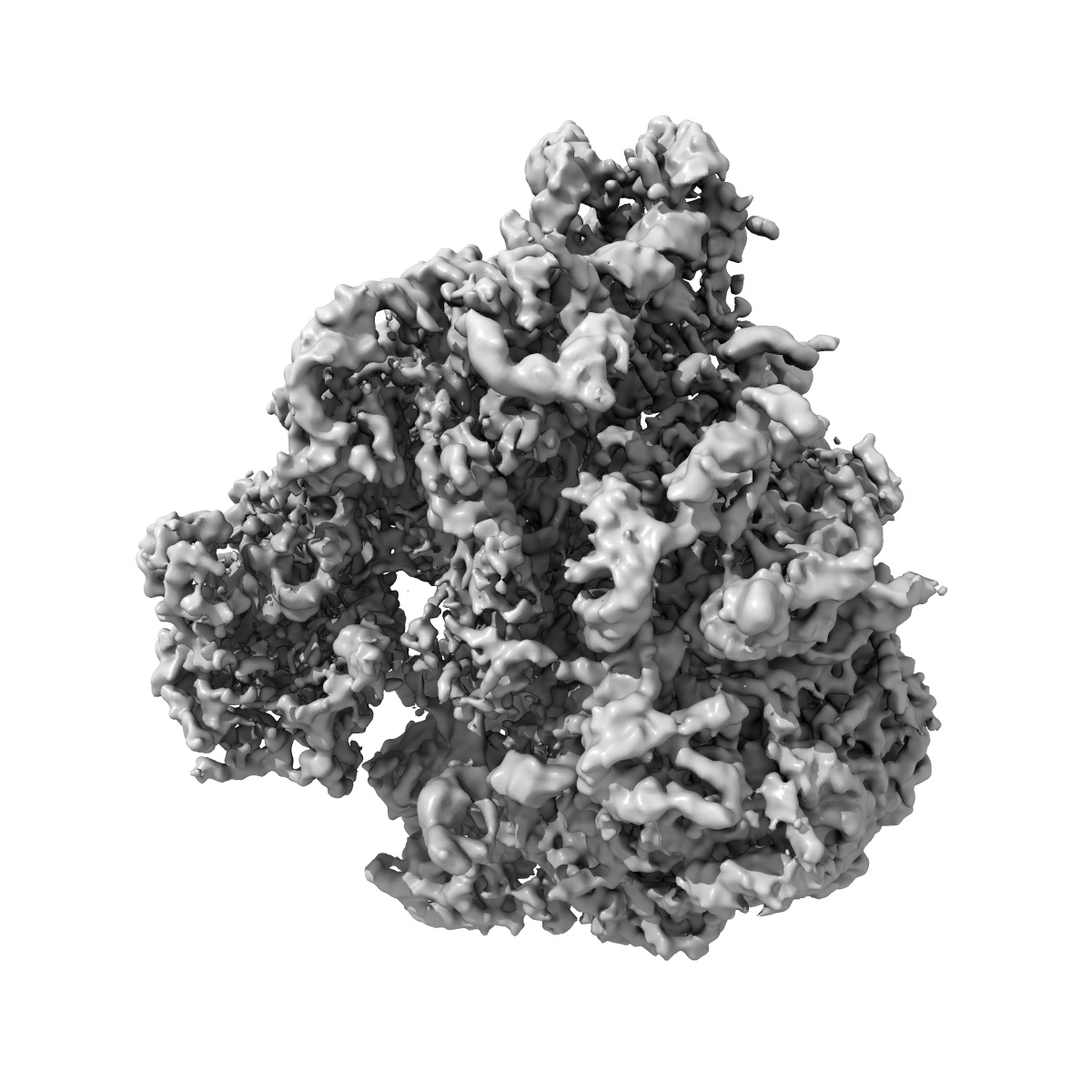
EMD-2239
Cryo-electron microscopy structure of the Trypanosoma brucei 80S ribosome
EMD-2239
Single-particle5.57 Å
 Deposition: 06/12/2012
Map released: 13/02/2013
Last modified: 27/02/2013
Concentration: 0.105
mg/mL
Buffer
pH: 7.2
Details: 20 mM Tris pH 7.2, 100mM MgCl2, 500 mM KCl, 5 mM beta-mercaptoethanol
Details: 20 mM Tris pH 7.2, 100mM MgCl2, 500 mM KCl, 5 mM beta-mercaptoethanol
Grid
Details: 300 mesh Copper/Molbydenum holey carbon-coated Quantifoil 2/4 grid (Quantifoil Micro Tools GmbH) containing an additional continuous thin layer of carbon
Vitrification
Cryogen name: ETHANE
Chamber humidity: 100%
Chamber temperature: 100 K
Instrument: FEI VITROBOT MARK IV
Method: Wait 30 sec, Blot 6 seconds, plunge
Chamber humidity: 100%
Chamber temperature: 100 K
Instrument: FEI VITROBOT MARK IV
Method: Wait 30 sec, Blot 6 seconds, plunge
Microscope: FEI POLARA 300
Illumination mode: FLOOD BEAM
Imaging mode: BRIGHT FIELD
Electron source: FIELD EMISSION GUN
Acceleration voltage: 300 kV
Nominal CS: 2.26 mm
Nominal defocus: 1.5 µm - 4.0 µm
Nominal magnification: 59000.0
Specimen holder model: SIDE ENTRY, EUCENTRIC
Illumination mode: FLOOD BEAM
Imaging mode: BRIGHT FIELD
Electron source: FIELD EMISSION GUN
Acceleration voltage: 300 kV
Nominal CS: 2.26 mm
Nominal defocus: 1.5 µm - 4.0 µm
Nominal magnification: 59000.0
Specimen holder model: SIDE ENTRY, EUCENTRIC
Image Recording
[1]
Detector category:
FILM
Detector model: KODAK SO-163 FILM
Scanner: NIKON SUPER COOLSCAN 9000
Number of real images: 1000
Average electron dose per image: 25 e/Å2
Bits per pixel: 32.0
Detector model: KODAK SO-163 FILM
Scanner: NIKON SUPER COOLSCAN 9000
Number of real images: 1000
Average electron dose per image: 25 e/Å2
Bits per pixel: 32.0
Details: Data were processed using SPIDER. The particles windows were automatically extracted from 1000 film-recorded micrographs and inspected manually. Standard SPIDER protocols for reference-based reconstruction, except that contrast transfer function (CTF) of the reconstructions was corrected by phase-flipping the particles using the defocus value estimated for each micrograph and a single reconstruction was obtained from the entire dataset using conjugate gradients with regularization (BP CG in SPIDER).
Final
reconstruction
Resolution: 5.57
Å
(
BY AUTHOR)
Resolution method: FSC 0.5 CUT-OFF
Number of images used: 164000
Algorithm: OTHER
Details:
Resolution method: FSC 0.5 CUT-OFF
Number of images used: 164000
Algorithm: OTHER
Details:
⌯ Applied Symmetry
Point group:
C1
Software
[1]
| Name | Version | Details |
|---|---|---|
| Spider | - | - |
CTF correction
Details:Phase-flip on each particle
Format: CCP4
Data type: IMAGE STORED AS FLOATING POINT NUMBER (4 BYTES)
Annotation details: Trypanosoma Brucei 80S Ribosome cryo-em reconstruction filtered according to the local FSC at 0.5
Details: ::::EMDATABANK.org::::EMD-2239::::
Data type: IMAGE STORED AS FLOATING POINT NUMBER (4 BYTES)
Annotation details: Trypanosoma Brucei 80S Ribosome cryo-em reconstruction filtered according to the local FSC at 0.5
Details: ::::EMDATABANK.org::::EMD-2239::::
⬡ Geometry
| X | Y | Z | |
|---|---|---|---|
| Dimensions | 359 | 359 | 359 |
| Origin | -179 | -179 | -179 |
| Spacing | 359 | 359 | 359 |
| Voxel size | 1.09 Å | 1.09 Å | 1.09 Å |
Contour list
| Primary | Level | Source |
|---|---|---|
| True | 108000.0 | AUTHOR |