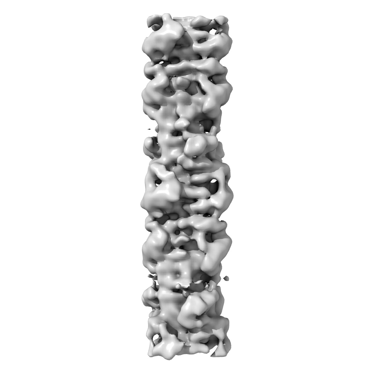
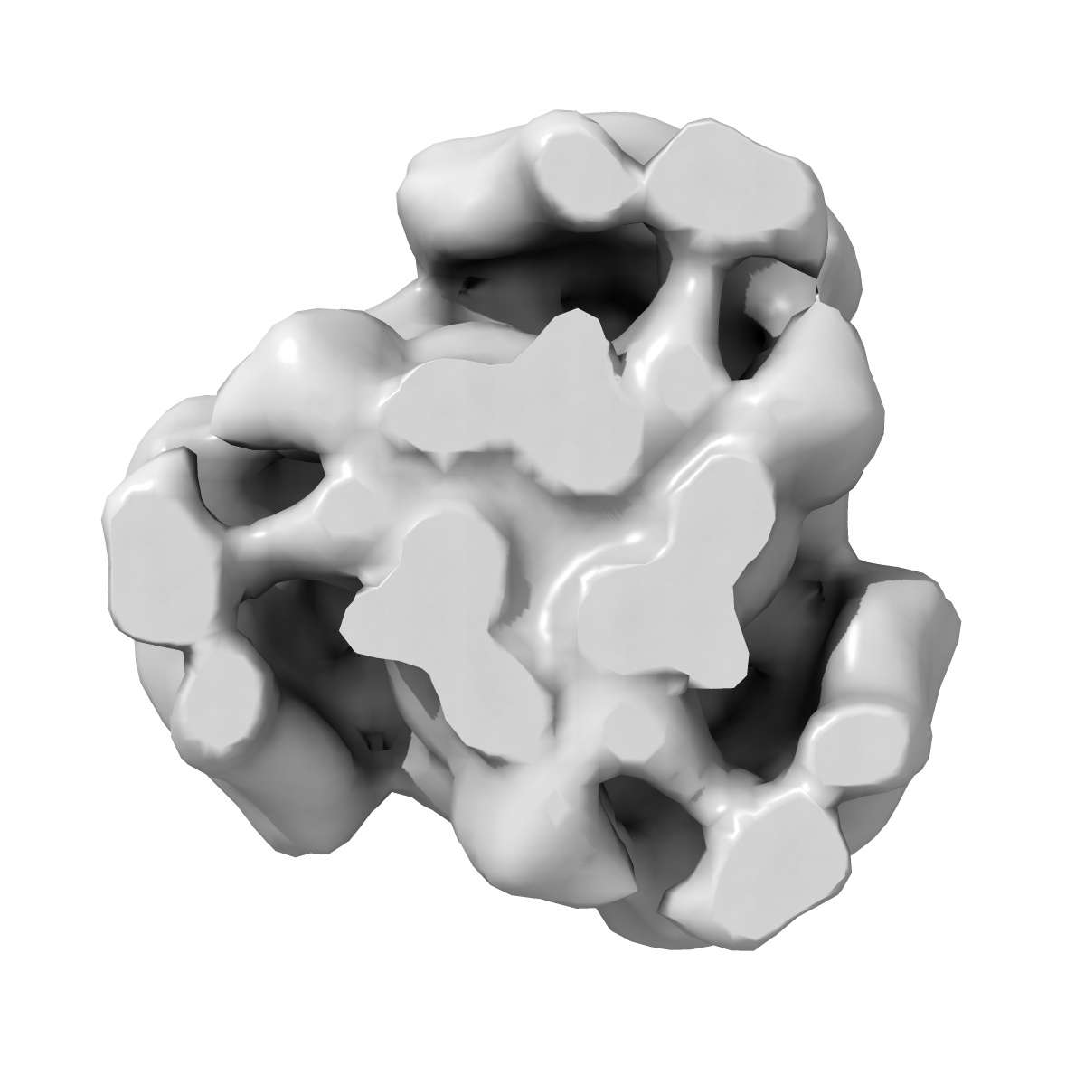
EMD-2240
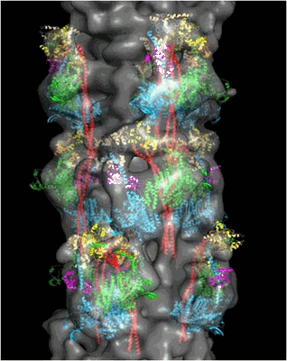
3D structure of myosin filaments isolated from human heart muscles by negative stain electron microscopy and single particle image analysis.
EMD-2240
Subtomogram averaging28.0 Å
 Deposition: 07/12/2012
Map released: 26/12/2012
Last modified: 26/12/2012
Method: Subtomogram averaging
Microscope: FEI TECNAI 20
Illumination mode: SPOT SCAN
Imaging mode: OTHER
Electron source: FIELD EMISSION GUN
Acceleration voltage: 200 kV
Nominal magnification: 29000.0
Specimen holder model: OTHER
Illumination mode: SPOT SCAN
Imaging mode: OTHER
Electron source: FIELD EMISSION GUN
Acceleration voltage: 200 kV
Nominal magnification: 29000.0
Specimen holder model: OTHER
Image Recording
[1]
Detector category:
CCD
Detector model: TVIPS TEMCAM-F415 (4k x 4k)
Sampling interval: 17 µm
Number of real images: 237
Average electron dose per image: 100 e/Å2
Bits per pixel: 16.0
Detector model: TVIPS TEMCAM-F415 (4k x 4k)
Sampling interval: 17 µm
Number of real images: 237
Average electron dose per image: 100 e/Å2
Bits per pixel: 16.0
Tilt Series
[1]
| Axis 1 | Axis 2 | |||||
|---|---|---|---|---|---|---|
| Min. | Max. | Inc. | Min. | Max. | Inc. | Rotation |
| - | - | - | - | - | - | - |
Format: CCP4
Data type: IMAGE STORED AS FLOATING POINT NUMBER (4 BYTES)
Annotation details: 3D map of human cardiac myosin filament
Details: ::::EMDATABANK.org::::EMD-2240::::
Data type: IMAGE STORED AS FLOATING POINT NUMBER (4 BYTES)
Annotation details: 3D map of human cardiac myosin filament
Details: ::::EMDATABANK.org::::EMD-2240::::
⬡ Geometry
| X | Y | Z | |
|---|---|---|---|
| Dimensions | 160 | 160 | 160 |
| Origin | -80 | -79 | -80 |
| Spacing | 160 | 160 | 160 |
| Voxel size | 5.94 Å | 5.94 Å | 5.94 Å |
Contour list
| Primary | Level | Source |
|---|---|---|
| True | 1.5 | AUTHOR |