EMD-2244
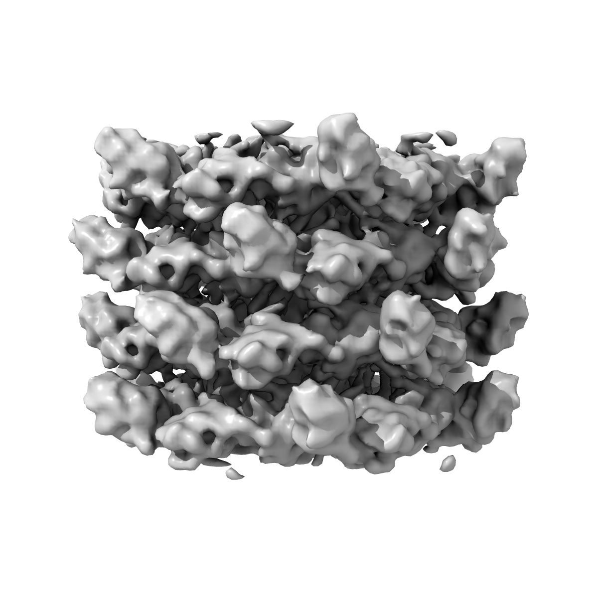
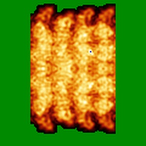
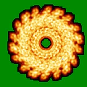
Cryo-electron microscopy of phirsl1 jumbo phage
EMD-2244
Helical reconstruction9.6 Å
 Deposition: 18/12/2012
Map released: 20/02/2013
Last modified: 20/04/2016
Buffer
pH: 7.5
Details: 50 mM Tris-HCl [pH 7.5], 100 mM NaCl, 10 mM MgSO4
Details: 50 mM Tris-HCl [pH 7.5], 100 mM NaCl, 10 mM MgSO4
Staining
Type:
NEGATIVE
Details: Four microliters of the bacteriophage sample (~0.1 mg/ml) were loaded between the mica-carbon interface. The sample was stained using 2% ammonium molybdate pH 7.5 and air-dried.
Details: Four microliters of the bacteriophage sample (~0.1 mg/ml) were loaded between the mica-carbon interface. The sample was stained using 2% ammonium molybdate pH 7.5 and air-dried.
Vitrification
Microscope: FEI POLARA 300
Illumination mode: SPOT SCAN
Imaging mode: BRIGHT FIELD
Electron source: FIELD EMISSION GUN
Acceleration voltage: 300 kV
Nominal CS: 2.3 mm
Nominal defocus: 1.355 µm - 5.295 µm
Nominal magnification: 31000.0
Specimen holder model: GATAN LIQUID NITROGEN
Alignment procedure: LEGACY (Astigmatism: Objective lens astigmatism was corrected at 59000, Electron beam tilt params: )
Image Recording
[1]
Detector category:
FILM
Detector model: KODAK SO-163 FILM
Scanner: ZEISS SCAI
Sampling interval: 7 µm
Number of real images: 52
Average electron dose per image: 20 e/Å2
Bits per pixel: 8.0
Detector model: KODAK SO-163 FILM
Scanner: ZEISS SCAI
Sampling interval: 7 µm
Number of real images: 52
Average electron dose per image: 20 e/Å2
Bits per pixel: 8.0
Details: Projection matching was done with SPIDER. IHRSR was used for helical parameters search and application.
Final
reconstruction
Resolution: 9.6
Å
(
BY AUTHOR)
Resolution method: FSC 0.5 CUT-OFF
Algorithm: OTHER
Details:
Resolution method: FSC 0.5 CUT-OFF
Algorithm: OTHER
Details:
⌯ Applied Symmetry
Software
[1]
| Name | Version | Details |
|---|---|---|
| SPIDER, IHRSR | - | - |
CTF correction
Details:Each particle
Format: CCP4
Data type: IMAGE STORED AS FLOATING POINT NUMBER (4 BYTES)
Annotation details: Reconstruction of the helical trunk of phirsl1 tail. A Bfactor of -538 has been applied to the reconstruction as well as a soft-edged mask.
Details: ::::EMDATABANK.org::::EMD-2244::::
Data type: IMAGE STORED AS FLOATING POINT NUMBER (4 BYTES)
Annotation details: Reconstruction of the helical trunk of phirsl1 tail. A Bfactor of -538 has been applied to the reconstruction as well as a soft-edged mask.
Details: ::::EMDATABANK.org::::EMD-2244::::
⬡ Geometry
| X | Y | Z | |
|---|---|---|---|
| Dimensions | 120 | 120 | 120 |
| Origin | -3 | 0 | -5 |
| Spacing | 120 | 120 | 120 |
| Voxel size | 2.26 Å | 2.26 Å | 2.26 Å |
Contour list
| Primary | Level | Source |
|---|---|---|
| True | 0.007 | AUTHOR |