EMD-2254
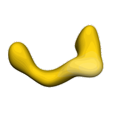
negative stain single-particle reconstruction of conformation VII of the Ltn1 E3 ubiquitin Ligase
EMD-2254
Single-particle53.4 Å
 Deposition: 25/12/2012
Map released: 16/01/2013
Last modified: 03/07/2013
Concentration: 0.01
mg/mL
Buffer
pH: 8.0
Details: 190 mM NaCl, 20 mM Tris, 1mM BME
Details: 190 mM NaCl, 20 mM Tris, 1mM BME
Staining
Type:
NEGATIVE
Details: 3 microliters of sample at a concentration of 0.01 mg/mL was applied to a C-flat grid with 2-micron-diameter holes overlaid by thin 1.5 nm continuous carbon. The specimen was stained with 2% uranyl formate 3 times, then let air-dry.
Details: 3 microliters of sample at a concentration of 0.01 mg/mL was applied to a C-flat grid with 2-micron-diameter holes overlaid by thin 1.5 nm continuous carbon. The specimen was stained with 2% uranyl formate 3 times, then let air-dry.
Grid
Details: C-flat grids overlaid with thin (~1.5 nm) carbon
Microscope: FEI TECNAI F20
Illumination mode: FLOOD BEAM
Imaging mode: BRIGHT FIELD
Electron source: FIELD EMISSION GUN
Acceleration voltage: 120 kV
Nominal CS: 2 mm
Nominal defocus: 1.2 µm - 2.6 µm
Nominal magnification: 50000.0
Calibrated magnification: 50000.0
Specimen holder model: SIDE ENTRY, EUCENTRIC
Specimen holder details: Single-tilt room temperature
Minimum tilt angle: -55
Maximum tilt angle: 0
Temperature
Average: 298
K
Image Recording
[1]
Detector category:
CCD
Detector model: GATAN ULTRASCAN 4000 (4k x 4k)
Number of real images: 489
Average electron dose per image: 15 e/Å2
Detector model: GATAN ULTRASCAN 4000 (4k x 4k)
Number of real images: 489
Average electron dose per image: 15 e/Å2
Details: Random-conical tilt reconstruction
Final
reconstruction
Resolution: 53.4
Å
(
BY AUTHOR)
Resolution method: FSC 0.5 CUT-OFF
Number of images used: 193
Algorithm: OTHER
Details:
Resolution method: FSC 0.5 CUT-OFF
Number of images used: 193
Algorithm: OTHER
Details:
⌯ Applied Symmetry
Point group:
C1
Software
[1]
| Name | Version | Details |
|---|---|---|
| Spider, Appion | - | - |
Final 2D classification
Number of classes:
1
CTF correction
Details:Each micrograph
Format: CCP4
Data type: IMAGE STORED AS FLOATING POINT NUMBER (4 BYTES)
Annotation details: Conformational snapshot VII of Ltn1
Details: ::::EMDATABANK.org::::EMD-2254::::
Data type: IMAGE STORED AS FLOATING POINT NUMBER (4 BYTES)
Annotation details: Conformational snapshot VII of Ltn1
Details: ::::EMDATABANK.org::::EMD-2254::::
⬡ Geometry
| X | Y | Z | |
|---|---|---|---|
| Dimensions | 160 | 160 | 160 |
| Origin | 0 | 0 | 0 |
| Spacing | 160 | 160 | 160 |
| Voxel size | 2.18 Å | 2.18 Å | 2.18 Å |
Contour list
| Primary | Level | Source |
|---|---|---|
| True | 0.8 | AUTHOR |