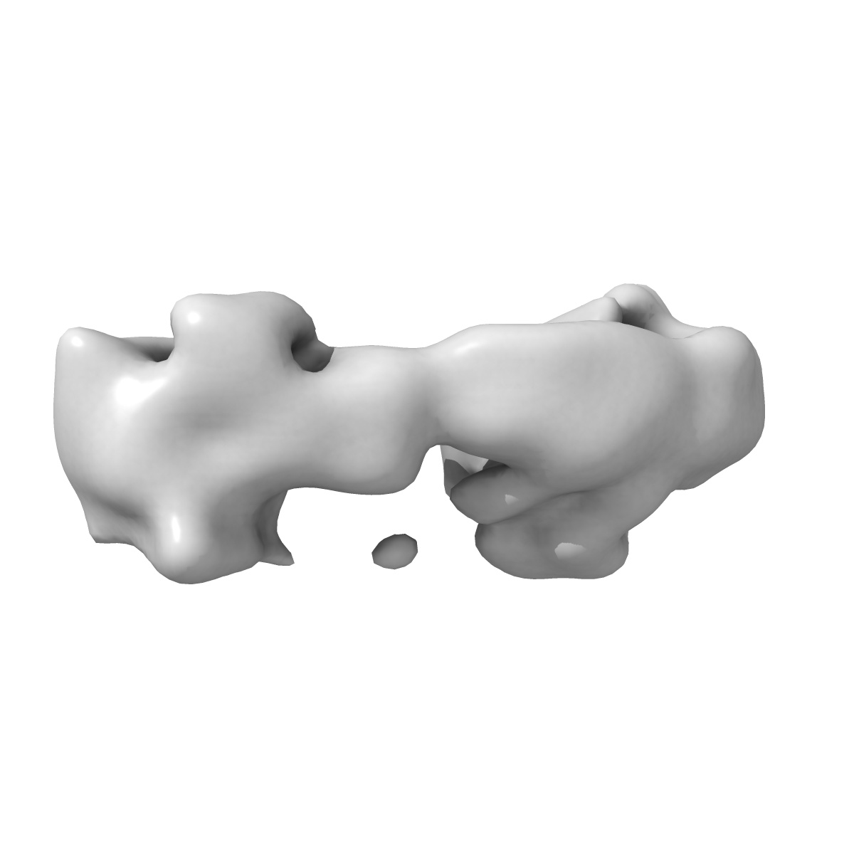
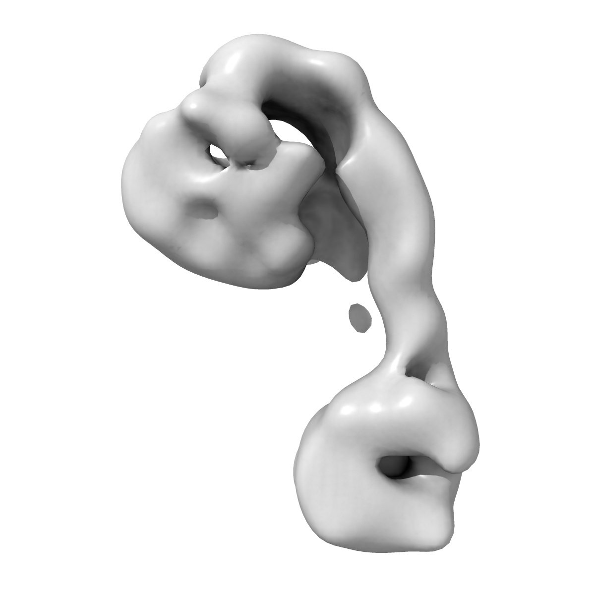
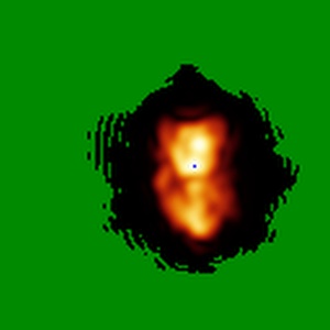
EMD-2280
Negative stain EM structure of the HOPS tethering complex (conformation 1)
EMD-2280
Single-particle30.4 Å
 Deposition: 16/01/2013
Map released: 23/01/2013
Last modified: 23/01/2013
Buffer
pH: 7.5
Details: 1 M NaCl, 50 mM Hepes/NaOH, 10% glycerol
Details: 1 M NaCl, 50 mM Hepes/NaOH, 10% glycerol
Staining
Type:
NEGATIVE
Details: Grids with adsorbed Protein floated on 0.07% Uranyl Formate for 1-3h at 4C.
Details: Grids with adsorbed Protein floated on 0.07% Uranyl Formate for 1-3h at 4C.
Grid
Details: Glow discharged 400 mesh copper grids with thin carbon support
Microscope: JEOL 1400
Illumination mode: FLOOD BEAM
Imaging mode: BRIGHT FIELD
Electron source: LAB6
Acceleration voltage: 120 kV
Nominal CS: 3.4 mm
Nominal defocus: 0.0013 µm - 0.0017 µm
Nominal magnification: 50000.0
Specimen holder model: JEOL
Illumination mode: FLOOD BEAM
Imaging mode: BRIGHT FIELD
Electron source: LAB6
Acceleration voltage: 120 kV
Nominal CS: 3.4 mm
Nominal defocus: 0.0013 µm - 0.0017 µm
Nominal magnification: 50000.0
Specimen holder model: JEOL
Image Recording
[1]
Detector category:
FILM
Detector model: KODAK SO-163 FILM
Scanner: OTHER
Sampling interval: 10.5 µm
Number of real images: 920
Average electron dose per image: 4.5 e/Å2
Bits per pixel: 16.0
Detector model: KODAK SO-163 FILM
Scanner: OTHER
Sampling interval: 10.5 µm
Number of real images: 920
Average electron dose per image: 4.5 e/Å2
Bits per pixel: 16.0
Final
reconstruction
Resolution: 30.4
Å
(
BY AUTHOR)
Resolution method: FSC 0.5 CUT-OFF
Number of images used: 2979
Algorithm: OTHER
Resolution method: FSC 0.5 CUT-OFF
Number of images used: 2979
Algorithm: OTHER
⌯ Applied Symmetry
Point group:
C1
Software
[1]
| Name | Version | Details |
|---|---|---|
| SPARX | - | - |
Format: CCP4
Data type: IMAGE STORED AS FLOATING POINT NUMBER (4 BYTES)
Annotation details: Reconstruction of HOPS tethering complex
Details: ::::EMDATABANK.org::::EMD-2280::::
Data type: IMAGE STORED AS FLOATING POINT NUMBER (4 BYTES)
Annotation details: Reconstruction of HOPS tethering complex
Details: ::::EMDATABANK.org::::EMD-2280::::
⬡ Geometry
| X | Y | Z | |
|---|---|---|---|
| Dimensions | 128 | 128 | 128 |
| Origin | 0 | 0 | 0 |
| Spacing | 128 | 128 | 128 |
| Voxel size | 4.2 Å | 4.2 Å | 4.2 Å |
Contour list
| Primary | Level | Source |
|---|---|---|
| True | 0.018 | AUTHOR |