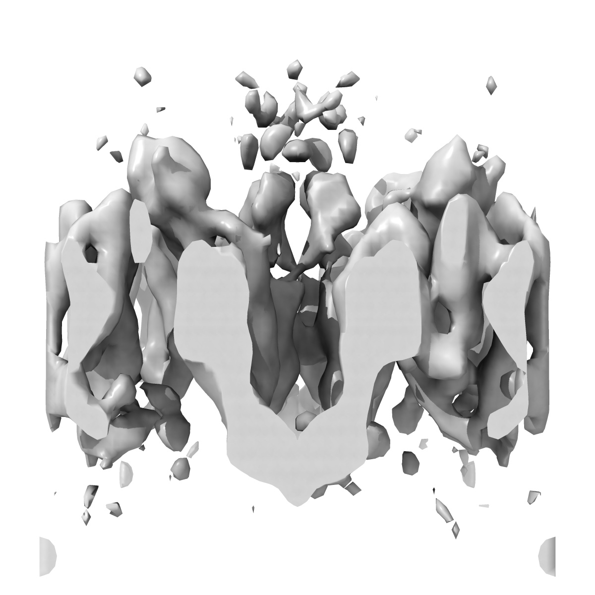
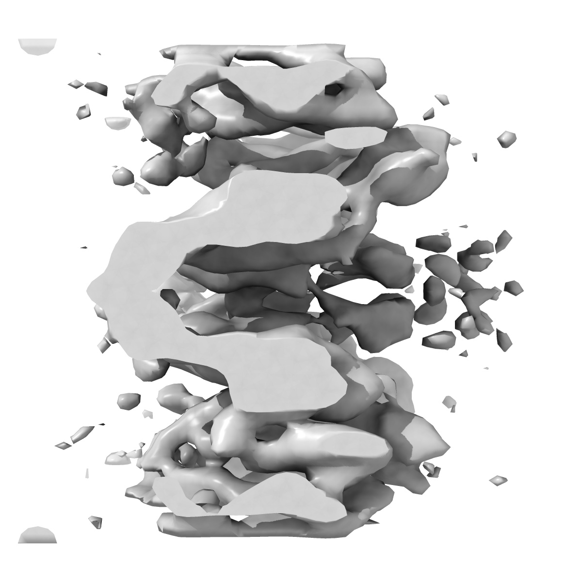
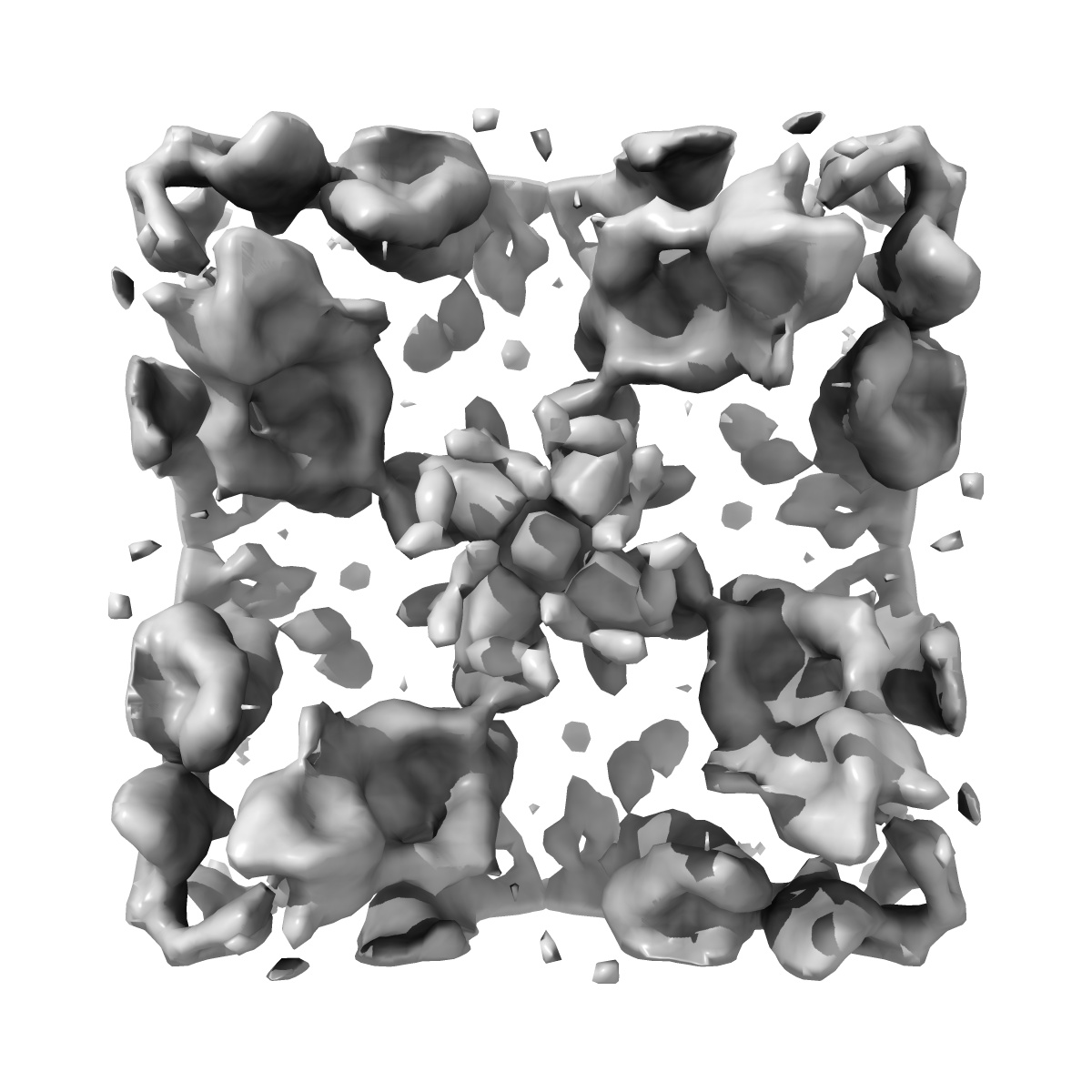
EMD-2347
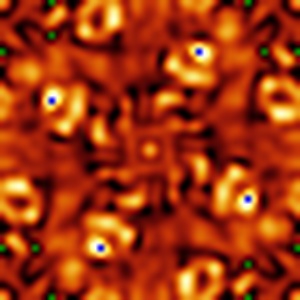
cryo-EM structure of the NavCt voltage-gated sodium channel
EMD-2347
Electron Crystallography9.0 Å
 Deposition: 28/03/2013
Map released: 10/07/2013
Last modified: 06/11/2013
Concentration: 4.0
mg/mL
Details: Crystals grown by dialysis
Details: Crystals grown by dialysis
Buffer
pH: 9.0
Details: 50mM glycine-NaOH pH9.0, 200mM NaCl, 4mM MgCl2, 5% glycerol, 5% methyl-2,4-pentanediol, 1.5mM NaN3
Details: 50mM glycine-NaOH pH9.0, 200mM NaCl, 4mM MgCl2, 5% glycerol, 5% methyl-2,4-pentanediol, 1.5mM NaN3
Grid
Details: Molybdenum EM grid
Vitrification
Crystal
formation
Details: Crystals grown by dialysis
Microscope: JEOL KYOTO-3000SFF
Illumination mode: FLOOD BEAM
Imaging mode: BRIGHT FIELD
Electron source: FIELD EMISSION GUN
Acceleration voltage: 300 kV
Nominal CS: 1.6 mm
Nominal defocus: 0.91 µm - 3.82 µm
Nominal magnification: 40000.0
Calibrated magnification: 39500.0
Specimen holder model: JEOL
Specimen holder details: Helium cooled, top entry
Minimum tilt angle: 0
Maximum tilt angle: 60
Temperature
Minimum: 4
K
Average: 4 K
Average: 4 K
Image Recording
[1]
Detector category:
FILM
Detector model: KODAK SO-163 FILM
Scanner: ZEISS SCAI
Sampling interval: 7 µm
Number of real images: 77
Average electron dose per image: 20 e/Å2
Bits per pixel: 14.0
Detector model: KODAK SO-163 FILM
Scanner: ZEISS SCAI
Sampling interval: 7 µm
Number of real images: 77
Average electron dose per image: 20 e/Å2
Bits per pixel: 14.0
Tilt Series
[1]
| Axis 1 | Axis 2 | |||||
|---|---|---|---|---|---|---|
| Min. | Max. | Inc. | Min. | Max. | Inc. | Rotation |
| 0 ° | 60 ° | - | - | - | - | - |
Details: Images were processed using MRC suite
Final
reconstruction
Crystal parameters
CTF correction
Details:Each micrographs
Format: CCP4
Data type: IMAGE STORED AS FLOATING POINT NUMBER (4 BYTES)
Annotation details: Cryo-EM structure of voltage-gated Na+ channel
Details: ::::EMDATABANK.org::::EMD-2347::::
Data type: IMAGE STORED AS FLOATING POINT NUMBER (4 BYTES)
Annotation details: Cryo-EM structure of voltage-gated Na+ channel
Details: ::::EMDATABANK.org::::EMD-2347::::
⬡ Geometry
| X | Y | Z | |
|---|---|---|---|
| Dimensions | 55 | 55 | 81 |
| Origin | 0 | 0 | -40 |
| Spacing | 55 | 55 | 81 |
| Voxel size | 2.090909 Å | 2.090909 Å | 2.2222223 Å |
Contour list
| Primary | Level | Source |
|---|---|---|
| True | 1.3 | EMDB |