EMD-2364
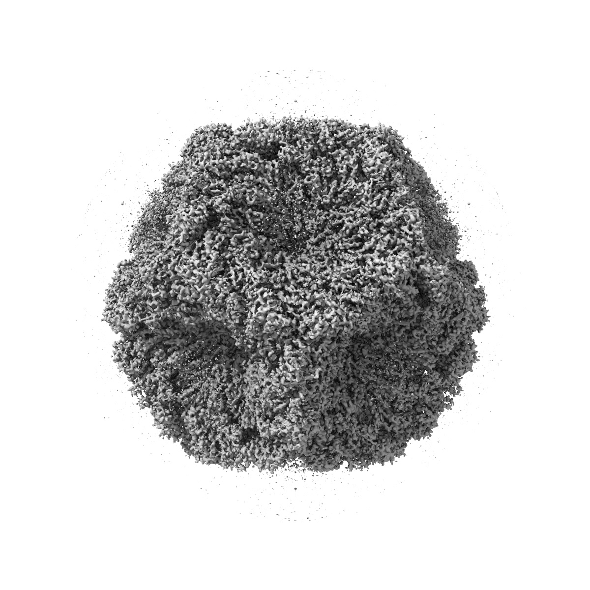
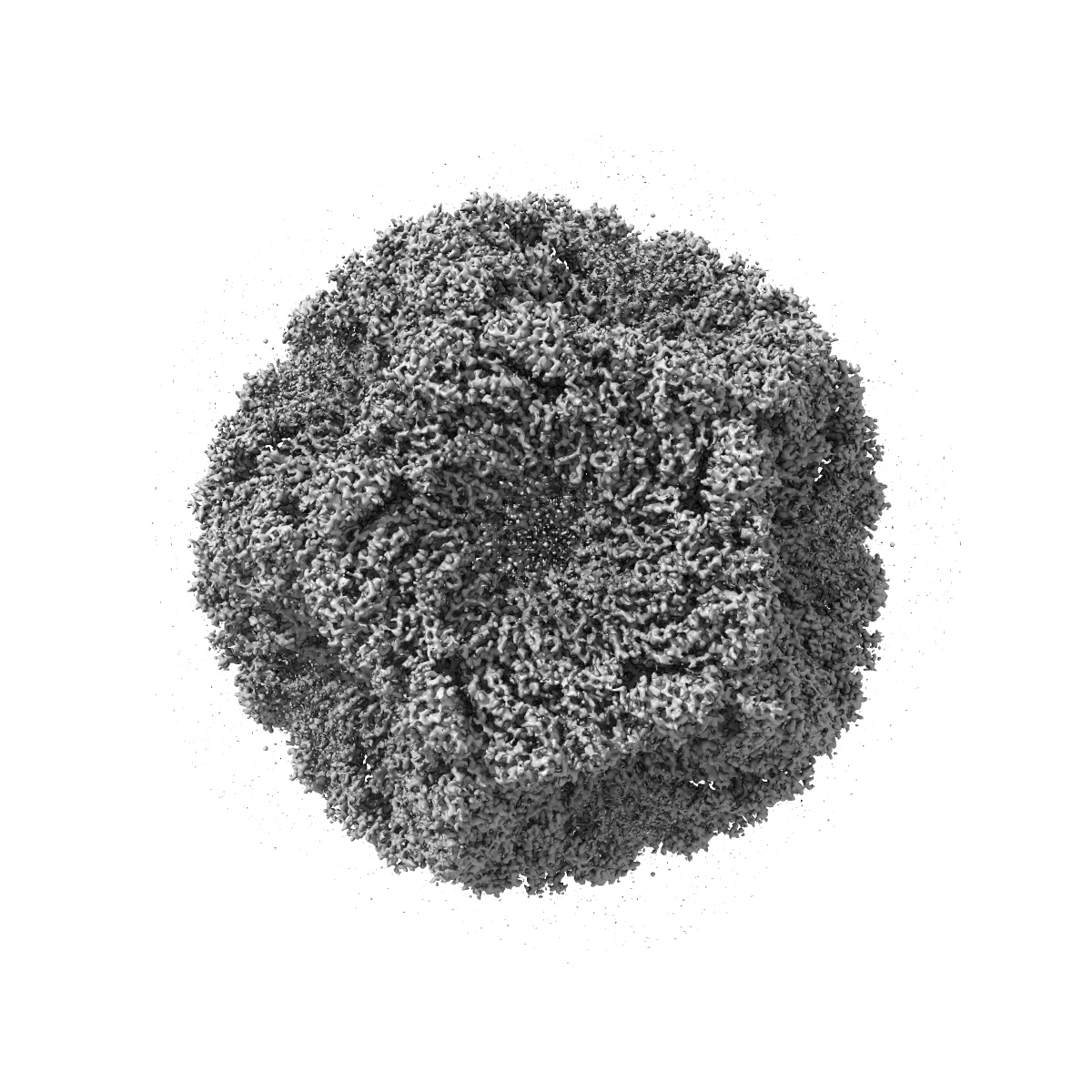
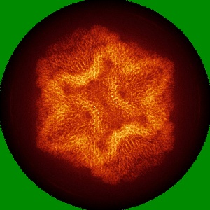
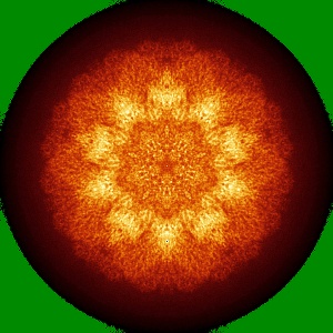
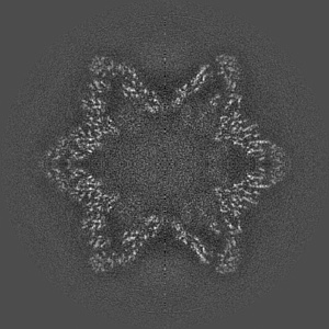
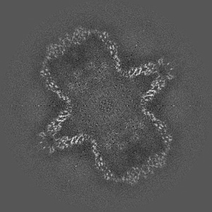
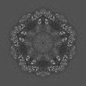
CryoEM reconstruction of the bacteriophage phi6 procapsid to the near-atomic resolution
EMD-2364
Single-particle4.4 Å
 Deposition: 17/04/2013
Map released: 14/08/2013
Last modified: 21/08/2013
Concentration: 10
mg/mL
Buffer
pH: 8.0
Details: 10 mM potassium phosphate, 5 mM MgCl2
Details: 10 mM potassium phosphate, 5 mM MgCl2
Grid
Details: 400 mesh C-flat holey carbon grid
Vitrification
Cryogen name: ETHANE
Chamber humidity: 80%
Chamber temperature: 100 K
Instrument: FEI VITROBOT MARK I
Method: Blot for 2 seconds before plunging.
Chamber humidity: 80%
Chamber temperature: 100 K
Instrument: FEI VITROBOT MARK I
Method: Blot for 2 seconds before plunging.
Microscope: FEI TITAN KRIOS
Illumination mode: FLOOD BEAM
Imaging mode: BRIGHT FIELD
Electron source: FIELD EMISSION GUN
Acceleration voltage: 300 kV
Nominal CS: 2.7 mm
Nominal defocus: 0.8 µm - 2.2 µm
Nominal magnification: 46000.0
Calibrated magnification: 44739.0
Specimen holder model: FEI TITAN KRIOS AUTOGRID HOLDER
Minimum tilt angle: 0
Maximum tilt angle: 0
Image Recording
[1]
Detector category:
FILM
Detector model: KODAK SO-163 FILM
Scanner: NIKON SUPER COOLSCAN 9000
Sampling interval: 6.35 µm
Number of real images: 220
Average electron dose per image: 15 e/Å2
Bits per pixel: 16.0
Detector model: KODAK SO-163 FILM
Scanner: NIKON SUPER COOLSCAN 9000
Sampling interval: 6.35 µm
Number of real images: 220
Average electron dose per image: 15 e/Å2
Bits per pixel: 16.0
Details: The particles were selected using e2boxer (EMAN) and manually pruned in bshow (BSOFT). The initial model was taken from our previous reconstruction at 7A resolution (EMD-2341). The final structure was reconstructed in EMAN.
Final
reconstruction
Resolution: 4.4
Å
(
BY AUTHOR)
Resolution method: FSC 0.5 CUT-OFF
Number of images used: 18326
Details:
Resolution method: FSC 0.5 CUT-OFF
Number of images used: 18326
Details:
⌯ Applied Symmetry
Point group:
I
Software
[1]
| Name | Version | Details |
|---|---|---|
| EMAN, BSOFT | - | - |
CTF correction
Details:Particles from each micrograph
Format: CCP4
Data type: IMAGE STORED AS FLOATING POINT NUMBER (4 BYTES)
Annotation details: Reconstruction of the wildtype P1247 procapsid of bacteriophage phi6
Details: ::::EMDATABANK.org::::EMD-2364::::
Data type: IMAGE STORED AS FLOATING POINT NUMBER (4 BYTES)
Annotation details: Reconstruction of the wildtype P1247 procapsid of bacteriophage phi6
Details: ::::EMDATABANK.org::::EMD-2364::::
⬡ Geometry
| X | Y | Z | |
|---|---|---|---|
| Dimensions | 420 | 420 | 420 |
| Origin | -210 | -210 | -210 |
| Spacing | 420 | 420 | 420 |
| Voxel size | 1.397 Å | 1.397 Å | 1.397 Å |
Contour list
| Primary | Level | Source |
|---|---|---|
| True | 2.0 | AUTHOR |