EMD-2380
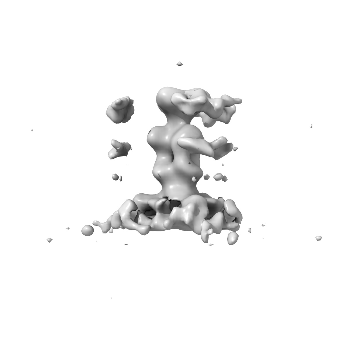
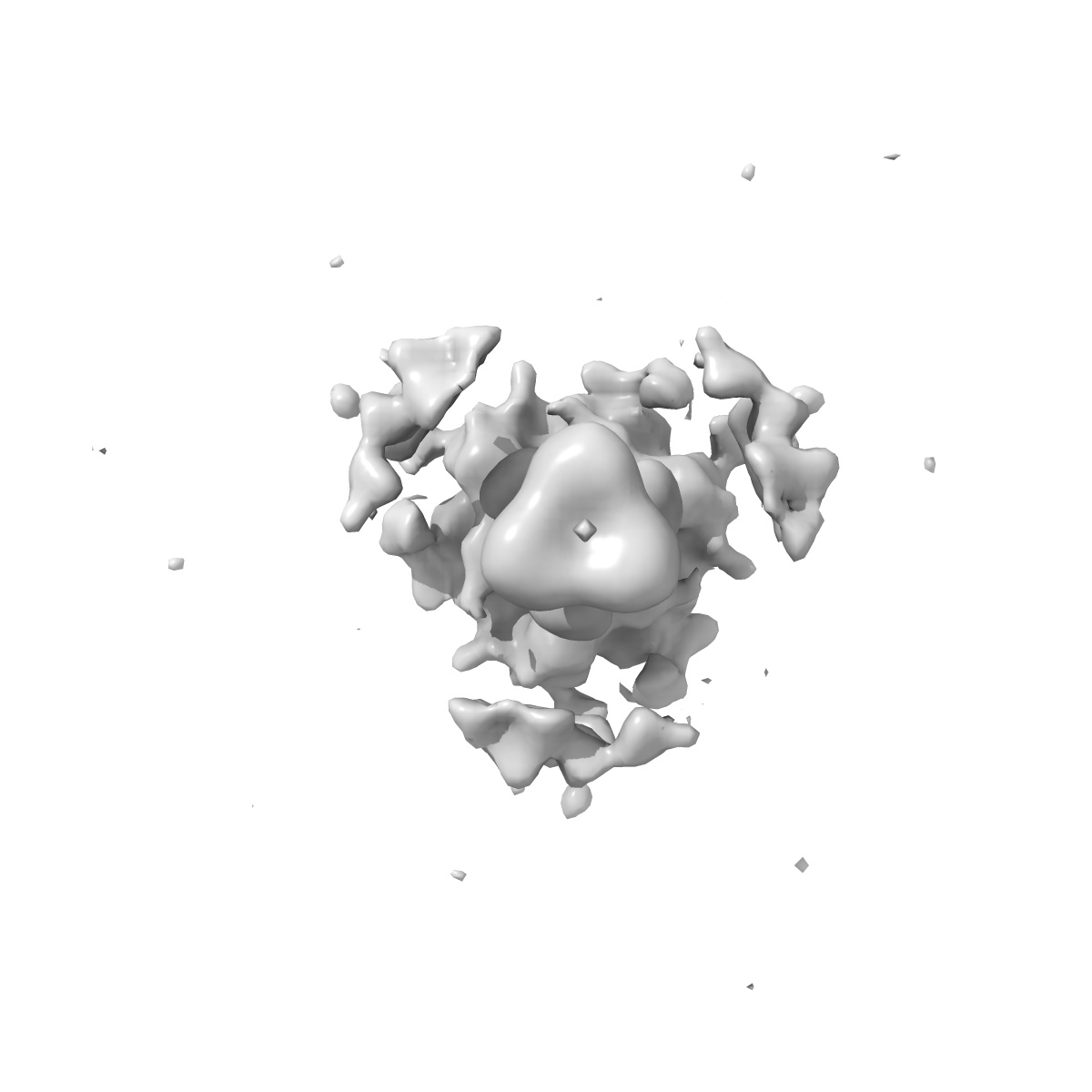
Structure of herpesvirus fusion glycoprotein B-bilayer complex revealing the protein-membrane and lateral protein-protein interaction
EMD-2380
Subtomogram averaging27.0 Å
 Deposition: 21/05/2013
Map released: 31/07/2013
Last modified: 21/08/2013
Concentration: 1.0
mg/mL
Buffer
pH: 5.5
Details: PBS with sodium citrate
Details: PBS with sodium citrate
Grid
Details: Cflat
Vitrification
Cryogen name: ETHANE-PROPANE MIXTURE
Chamber temperature: 120 K
Instrument: OTHER
Method: Blot manually for 3 s before plunging
Chamber temperature: 120 K
Instrument: OTHER
Method: Blot manually for 3 s before plunging
Microscope: FEI TECNAI F20
Illumination mode: FLOOD BEAM
Imaging mode: BRIGHT FIELD
Electron source: FIELD EMISSION GUN
Acceleration voltage: 200 kV
Nominal CS: 2 mm
Nominal defocus: 2.0 µm - 2.0 µm
Calibrated magnification: 67000.0
Specimen holder model: SIDE ENTRY, EUCENTRIC
Specimen holder details: Liquid nitrogen cooled
Illumination mode: FLOOD BEAM
Imaging mode: BRIGHT FIELD
Electron source: FIELD EMISSION GUN
Acceleration voltage: 200 kV
Nominal CS: 2 mm
Nominal defocus: 2.0 µm - 2.0 µm
Calibrated magnification: 67000.0
Specimen holder model: SIDE ENTRY, EUCENTRIC
Specimen holder details: Liquid nitrogen cooled
Image Recording
[1]
Detector category:
CCD
Detector model: GATAN ULTRASCAN 4000 (4k x 4k)
Sampling interval: 15 µm
Number of real images: 9
Average electron dose per image: 100 e/Å2
Bits per pixel: 12.0
Details: The dataset consists of 9 tomograms (containing 38 liposomes with bound gB). Data were binned by factor of 2.
Detector model: GATAN ULTRASCAN 4000 (4k x 4k)
Sampling interval: 15 µm
Number of real images: 9
Average electron dose per image: 100 e/Å2
Bits per pixel: 12.0
Details: The dataset consists of 9 tomograms (containing 38 liposomes with bound gB). Data were binned by factor of 2.
Tilt Series
[1]
| Axis 1 | Axis 2 | |||||
|---|---|---|---|---|---|---|
| Min. | Max. | Inc. | Min. | Max. | Inc. | Rotation |
| -60 ° | 60 ° | - | - | - | - | - |
Details: The sub-tomograms were picked manually from tomographic reconstructions of 38 liposomes
Final
reconstruction
Resolution: 27.0
Å
(
BY AUTHOR)
Resolution method: FSC 0.5 CUT-OFF
Algorithm: OTHER
Details: The best 786 spikes (of 996) were selected based on constrained cross correlation coefficient and by excluding overlaps. All three Euler angles of the spike were refined.
Resolution method: FSC 0.5 CUT-OFF
Algorithm: OTHER
Details: The best 786 spikes (of 996) were selected based on constrained cross correlation coefficient and by excluding overlaps. All three Euler angles of the spike were refined.
⌯ Applied Symmetry
Point group:
C3
Software
[1]
| Name | Version | Details |
|---|---|---|
| Jsubtomo | - | - |
CTF correction
Details:Low pass filter to the first zero crossing of the CTF
Format: CCP4
Data type: IMAGE STORED AS FLOATING POINT NUMBER (4 BYTES)
Annotation details: Subtomogram average of HSV-1 glycoprotein B bound to a lipid bilayer
Details: ::::EMDATABANK.org::::EMD-2380::::
Data type: IMAGE STORED AS FLOATING POINT NUMBER (4 BYTES)
Annotation details: Subtomogram average of HSV-1 glycoprotein B bound to a lipid bilayer
Details: ::::EMDATABANK.org::::EMD-2380::::
⬡ Geometry
| X | Y | Z | |
|---|---|---|---|
| Dimensions | 100 | 100 | 100 |
| Origin | -50 | -50 | -50 |
| Spacing | 100 | 100 | 100 |
| Voxel size | 4.6 Å | 4.6 Å | 4.6 Å |
Contour list
| Primary | Level | Source |
|---|---|---|
| True | 2.0 | AUTHOR |