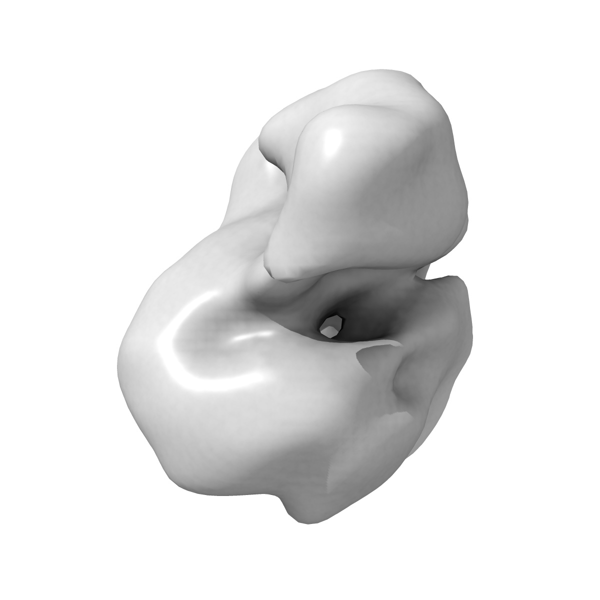
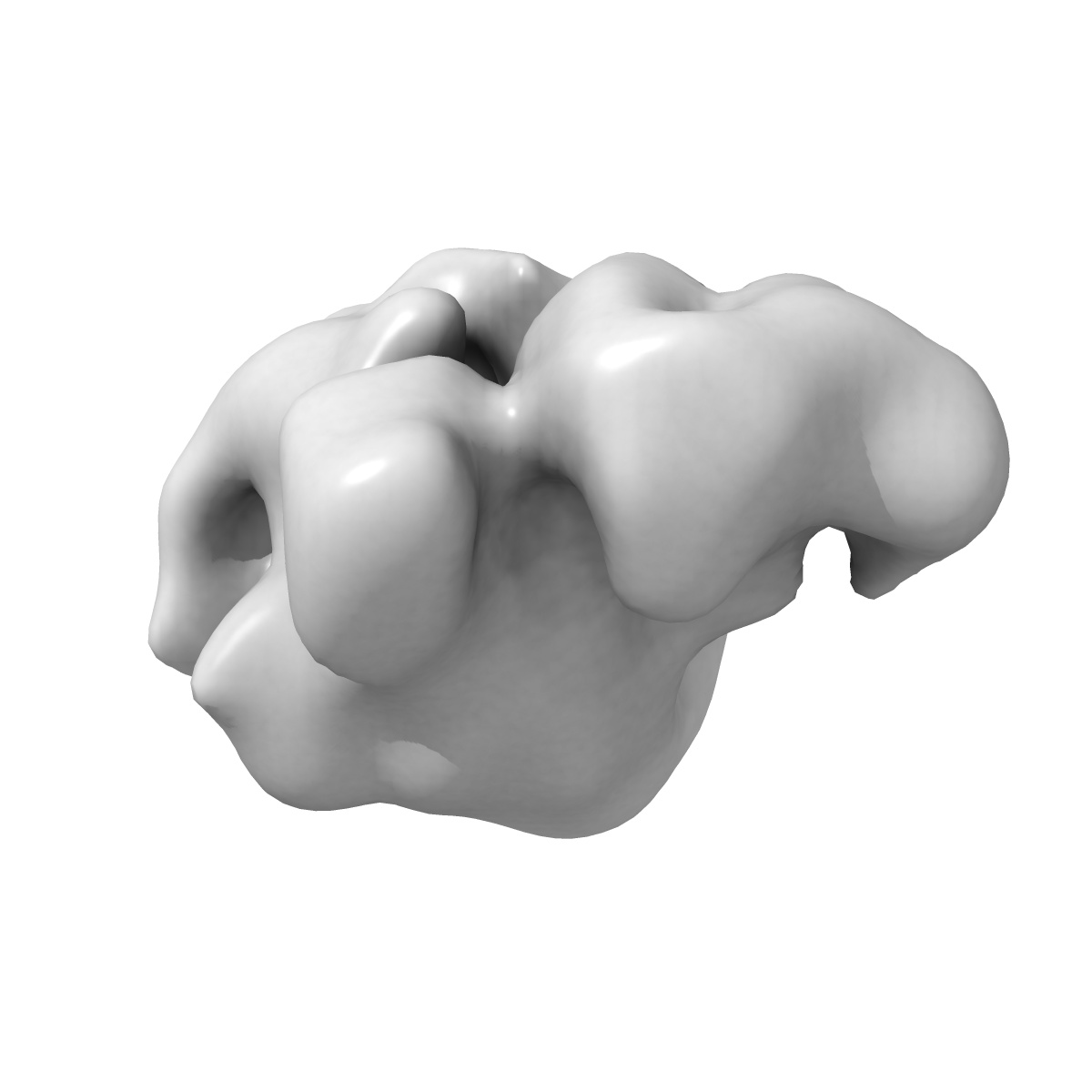
EMD-2502
Substrate Recruitment Pathways in the Yeast Exosome by Electron Microscopy
EMD-2502
Single-particle24.0 Å
 Deposition: 23/10/2013
Map released: 25/12/2013
Last modified: 12/10/2016
Concentration: 2
mg/mL
Buffer
pH: 8.0
Details: 150mM NaCl, 50mM Tris-HCL,1mM DTT, 2mM MgCl2
Details: 150mM NaCl, 50mM Tris-HCL,1mM DTT, 2mM MgCl2
Grid
Details: Quantifoil grids (2/2) with 2~3 nm thin carbon on top
Vitrification
Cryogen name: ETHANE
Chamber humidity: 100%
Chamber temperature: 80 K
Instrument: FEI VITROBOT MARK IV
Method: 4 ul of the reaction solution was then applied to glow-discharged C-flat grids (1.2/1.3) covered with a layer of continuous carbon with a thickness of ~4nm. The grids were then blotted and plunged into liquid ethane in a FEI Vitrobot Mark IV
Microscope: FEI TITAN KRIOS
Illumination mode: FLOOD BEAM
Imaging mode: BRIGHT FIELD
Electron source: FIELD EMISSION GUN
Acceleration voltage: 120 kV
Nominal CS: 2.7 mm
Nominal defocus: -0.5 µm - -3.5 µm
Nominal magnification: 59000.0
Calibrated magnification: 59000.0
Specimen holder model: FEI TITAN KRIOS AUTOGRID HOLDER
Illumination mode: FLOOD BEAM
Imaging mode: BRIGHT FIELD
Electron source: FIELD EMISSION GUN
Acceleration voltage: 120 kV
Nominal CS: 2.7 mm
Nominal defocus: -0.5 µm - -3.5 µm
Nominal magnification: 59000.0
Calibrated magnification: 59000.0
Specimen holder model: FEI TITAN KRIOS AUTOGRID HOLDER
Temperature
Minimum: 80
K
Average: 82 K
Maximum: 85 K
Average: 82 K
Maximum: 85 K
Image Recording
[1]
Detector category:
CCD
Detector model: GENERIC GATAN (4k x 4k)
Number of real images: 1200
Average electron dose per image: 20 e/Å2
Detector model: GENERIC GATAN (4k x 4k)
Number of real images: 1200
Average electron dose per image: 20 e/Å2
Details: Micrographs of RE were collected using the AutoEMation software (Lei and Frank, 2005) installed on the microscope at low-dose condition with a dose of ~20 electron/A2 and a defocus value ranging from -1.2 to -4 um. The micrographs were collected on a FEI Eagle CCD camera with a pixel size of 1.5 A.
Final
reconstruction
Resolution: 24.0
Å
(
BY AUTHOR)
Resolution method: FSC 0.5 CUT-OFF
Number of images used: 27200
Details:
Resolution method: FSC 0.5 CUT-OFF
Number of images used: 27200
Details:
⌯ Applied Symmetry
Point group:
C1
Software
[1]
| Name | Version | Details |
|---|---|---|
| Eman, imagic, relion | - | - |
⦩ Final angle assignment
Details: RELION: theta 45 degrees, phi 180 degrees
CTF correction
Details:Both each particle and micrograph
Format: CCP4
Data type: IMAGE STORED AS FLOATING POINT NUMBER (4 BYTES)
Annotation details: Apo-exosome
Details: ::::EMDATABANK.org::::EMD-2502::::
Data type: IMAGE STORED AS FLOATING POINT NUMBER (4 BYTES)
Annotation details: Apo-exosome
Details: ::::EMDATABANK.org::::EMD-2502::::
⬡ Geometry
| X | Y | Z | |
|---|---|---|---|
| Dimensions | 120 | 120 | 120 |
| Origin | 0 | 0 | 0 |
| Spacing | 120 | 120 | 120 |
| Voxel size | 3.0 Å | 3.0 Å | 3.0 Å |
Contour list
| Primary | Level | Source |
|---|---|---|
| True | 0.05 | AUTHOR |