EMD-5375
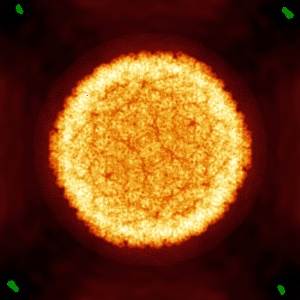
Direct electron detection yields cryo-EM reconstructions at resolutions beyond .75 Nyquist frequency
EMD-5375
Single-particle8.5 Å
 Deposition: 20/12/2011
Map released: 24/10/2012
Last modified: 24/10/2012
Concentration: 1
mg/mL
Buffer
pH: 7.6
Details: 50 mM Tris pH 7.6, 25 mM NaCl, 2mM EDTA
Details: 50 mM Tris pH 7.6, 25 mM NaCl, 2mM EDTA
Grid
Details: 400 mesh copper grid
Microscope: JEOL 2010F
Illumination mode: FLOOD BEAM
Imaging mode: BRIGHT FIELD
Electron source: FIELD EMISSION GUN
Acceleration voltage: 200 kV
Nominal CS: 2.0 mm
Nominal defocus: 2.0 µm - 5.0 µm
Nominal magnification: 15000.0
Calibrated magnification: 17200.0
Specimen holder model: GATAN LIQUID NITROGEN
Specimen holder details: Side Entry
Alignment procedure: LEGACY (Astigmatism: objective lens astigmatism was corrected at 100,000 times magnification, Electron beam tilt params: )
Temperature
Minimum: 100
K
Average: 100 K
Maximum: 100 K
Average: 100 K
Maximum: 100 K
Image Recording
[1]
Detector category:
CCD
Detector model: DIRECT ELECTRON DE-12 (4k x 3k)
Sampling interval: 6 µm
Number of real images: 200
Average electron dose per image: 17 e/Å2
Details: Images were collected on a CMOS type detector DE-12 from Direct Electron
Detector model: DIRECT ELECTRON DE-12 (4k x 3k)
Sampling interval: 6 µm
Number of real images: 200
Average electron dose per image: 17 e/Å2
Details: Images were collected on a CMOS type detector DE-12 from Direct Electron
Final
reconstruction
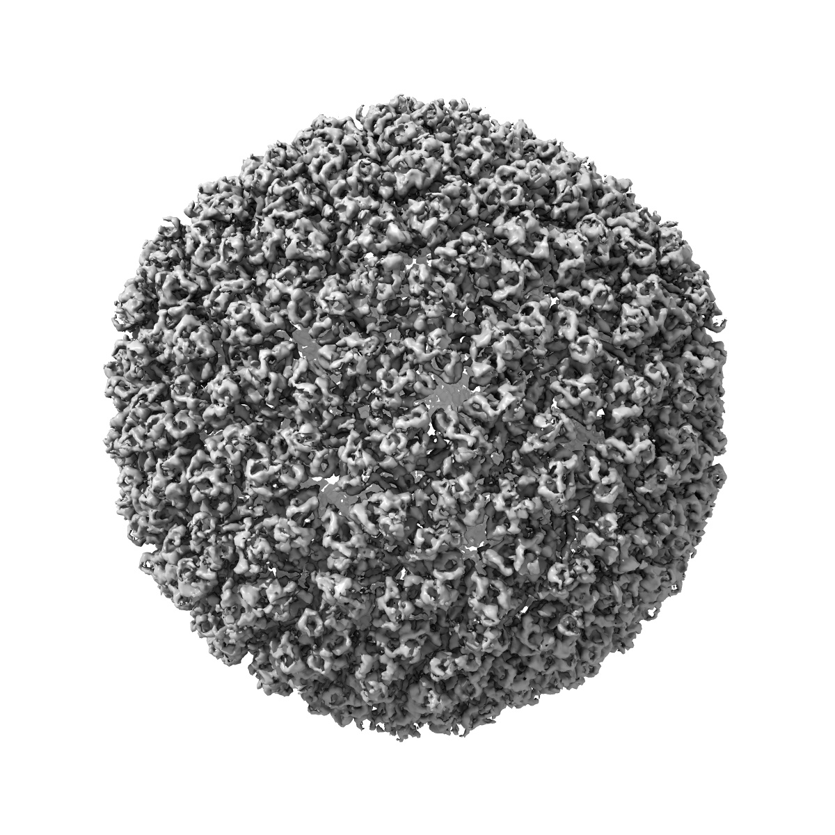
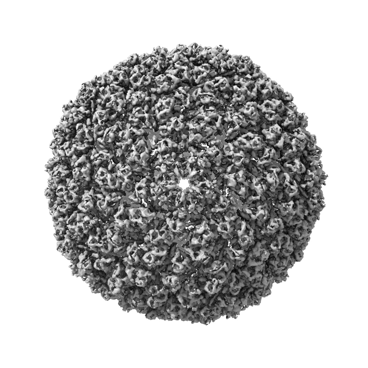
Resolution: 8.5
Å
(
BY AUTHOR)
Resolution method: FSC 0.5 CUT-OFF
Number of images used: 7500
Algorithm: OTHER
Resolution method: FSC 0.5 CUT-OFF
Number of images used: 7500
Algorithm: OTHER
⌯ Applied Symmetry
Point group:
I
Software
[1]
| Name | Version | Details |
|---|---|---|
| MPSA | - | - |
CTF correction
Details:Each Micrograph
Format: CCP4
Data type: IMAGE STORED AS FLOATING POINT NUMBER (4 BYTES)
Annotation details: Icosahedral reconstruction of bacteriophage P22
Details: ::::EMDATABANK.org::::EMD-5375::::
Data type: IMAGE STORED AS FLOATING POINT NUMBER (4 BYTES)
Annotation details: Icosahedral reconstruction of bacteriophage P22
Details: ::::EMDATABANK.org::::EMD-5375::::
⬡ Geometry
| X | Y | Z | |
|---|---|---|---|
| Dimensions | 384 | 384 | 384 |
| Origin | -128 | -128 | -128 |
| Spacing | 384 | 384 | 384 |
| Voxel size | 3.48 Å | 3.48 Å | 3.48 Å |
Contour list
| Primary | Level | Source |
|---|---|---|
| True | 0.8 | AUTHOR |