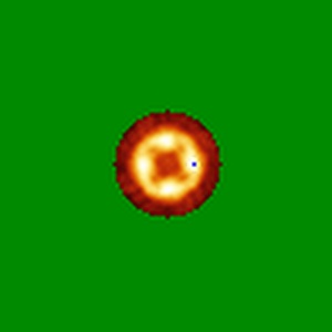
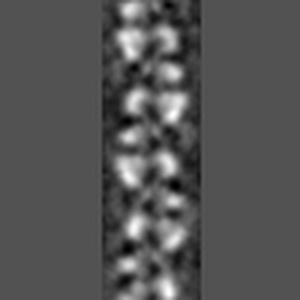
EMD-5444
MDA5-dsRNA filament
EMD-5444
Helical reconstruction20.0 Å
 Deposition: 04/07/2012
Map released: 24/10/2012
Last modified: 17/07/2013
Microscope: FEI TECNAI 12
Illumination mode: FLOOD BEAM
Imaging mode: BRIGHT FIELD
Electron source: TUNGSTEN HAIRPIN
Acceleration voltage: 80 kV
Specimen holder model: OTHER
Illumination mode: FLOOD BEAM
Imaging mode: BRIGHT FIELD
Electron source: TUNGSTEN HAIRPIN
Acceleration voltage: 80 kV
Specimen holder model: OTHER
Image Recording
[1]
Detector category:
FILM
Detector model: KODAK SO-163 FILM
Scanner: NIKON COOLSCAN
Sampling interval: 4.16 µm
Bits per pixel: 14.0
Detector model: KODAK SO-163 FILM
Scanner: NIKON COOLSCAN
Sampling interval: 4.16 µm
Bits per pixel: 14.0
Details: The particles were processed using IHRSR
Final
reconstruction
Resolution: 20.0
Å
(
BY AUTHOR)
Resolution method: FSC 0.5 CUT-OFF
Algorithm: OTHER
Details:
Resolution method: FSC 0.5 CUT-OFF
Algorithm: OTHER
Details:
⌯ Applied Symmetry
ΔΖ:
43.6
Å
ΔΦ: 82.7°
ΔΦ: 82.7°
Software
[1]
| Name | Version | Details |
|---|---|---|
| Spider | - | - |
Format: CCP4
Data type: IMAGE STORED AS FLOATING POINT NUMBER (4 BYTES)
Annotation details: Reconstruction of MDA5-dsRNA
Details: ::::EMDATABANK.org::::EMD-5444::::
Data type: IMAGE STORED AS FLOATING POINT NUMBER (4 BYTES)
Annotation details: Reconstruction of MDA5-dsRNA
Details: ::::EMDATABANK.org::::EMD-5444::::
⬡ Geometry
| X | Y | Z | |
|---|---|---|---|
| Dimensions | 100 | 100 | 100 |
| Origin | 0 | 0 | 0 |
| Spacing | 100 | 100 | 100 |
| Voxel size | 4.16 Å | 4.16 Å | 4.16 Å |
Contour list
| Primary | Level | Source |
|---|---|---|
| True | 0.175 | EMDB |