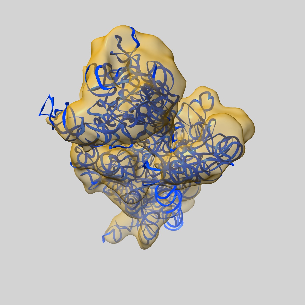
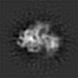
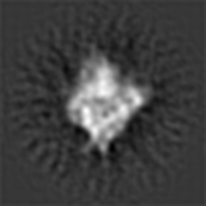
EMD-5509
Dissecting the in vivo assembly of the 30S ribosomal subunit reveals the role of RimM
EMD-5509
Single-particle16.5 Å
 Deposition: 28/09/2012
Map released: 16/01/2013
Last modified: 06/03/2013
Buffer
pH: 7.5
Details: 150 mM NH4Cl,10mM Tris-HCL,10mM MgCl2
Details: 150 mM NH4Cl,10mM Tris-HCL,10mM MgCl2
Vitrification
Cryogen name: ETHANE
Chamber humidity: 100%
Instrument: FEI VITROBOT MARK IV
Method: Blot for 1 seconds before plunging
Chamber humidity: 100%
Instrument: FEI VITROBOT MARK IV
Method: Blot for 1 seconds before plunging
Microscope: FEI TITAN KRIOS
Illumination mode: FLOOD BEAM
Imaging mode: BRIGHT FIELD
Electron source: FIELD EMISSION GUN
Acceleration voltage: 300 kV
Nominal defocus: 1.5 µm - 3.8 µm
Nominal magnification: 59000.0
Specimen holder model: GATAN LIQUID NITROGEN
Illumination mode: FLOOD BEAM
Imaging mode: BRIGHT FIELD
Electron source: FIELD EMISSION GUN
Acceleration voltage: 300 kV
Nominal defocus: 1.5 µm - 3.8 µm
Nominal magnification: 59000.0
Specimen holder model: GATAN LIQUID NITROGEN
Image Recording
[1]
Details: This is a classification volume (No. 4) using ML3D methods.
Final
reconstruction
Resolution: 16.5
Å
(
BY AUTHOR)
Resolution method: FSC 0.5 CUT-OFF
Number of images used: 25631
Algorithm: OTHER
Details:
Resolution method: FSC 0.5 CUT-OFF
Number of images used: 25631
Algorithm: OTHER
Details:
⌯ Applied Symmetry
Point group:
C1
Software
[1]
| Name | Version | Details |
|---|---|---|
| SPIDER | - | - |
CTF correction
Details:Weiner filter
Format: CCP4
Data type: IMAGE STORED AS FLOATING POINT NUMBER (4 BYTES)
Annotation details: The map has been normalized to N(0,1)
Details: ::::EMDATABANK.org::::EMD-5509::::
Data type: IMAGE STORED AS FLOATING POINT NUMBER (4 BYTES)
Annotation details: The map has been normalized to N(0,1)
Details: ::::EMDATABANK.org::::EMD-5509::::
⬡ Geometry
| X | Y | Z | |
|---|---|---|---|
| Dimensions | 125 | 125 | 125 |
| Origin | -62 | -62 | -62 |
| Spacing | 125 | 125 | 125 |
| Voxel size | 3.00 Å | 3.00 Å | 3.00 Å |
Contour list
| Primary | Level | Source |
|---|---|---|
| True | -2.3 | AUTHOR |