EMD-5529
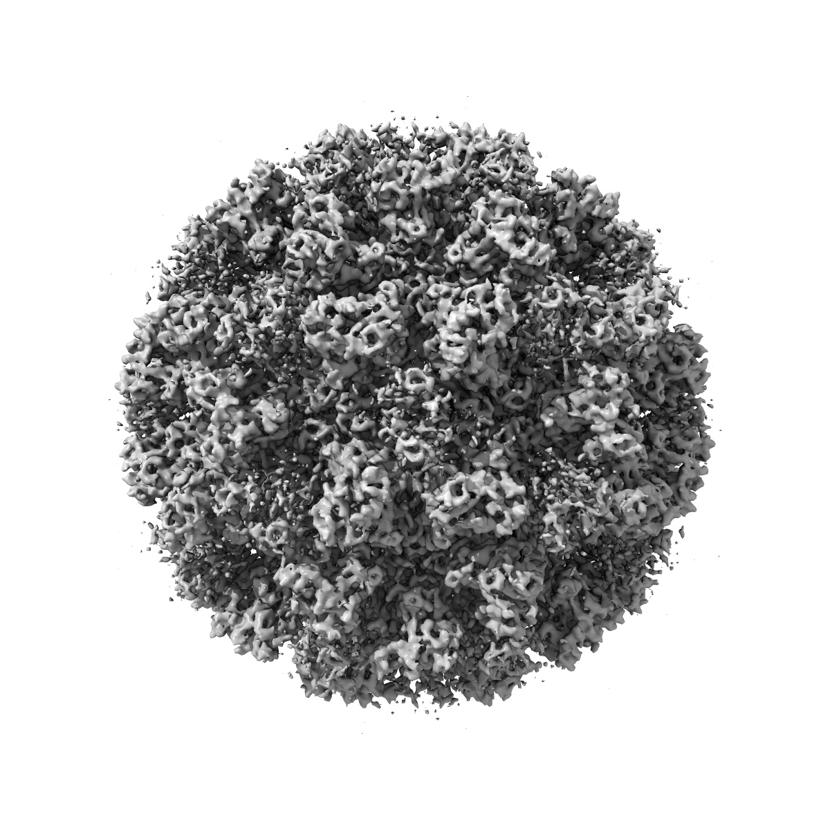
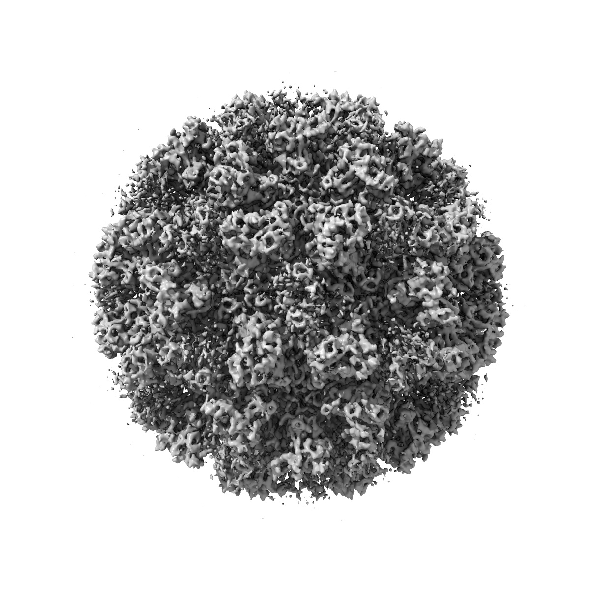
6.3 A Cryo-EM Structure of a Novel Calicivirus, Tulane Virus
EMD-5529
Single-particle6.3 Å
 Deposition: 30/11/2012
Map released: 03/04/2013
Last modified: 03/04/2013
Buffer
pH: 7.4
Details: 137mM NaCl, 2.7mM KCl, 10mM Na2HPO4, 2mM KH2PO4, pH 7.4
Details: 137mM NaCl, 2.7mM KCl, 10mM Na2HPO4, 2mM KH2PO4, pH 7.4
Staining
Type:
NEGATIVE
Details: Grids with sample floated on 2% uranyl acetate for 30 seconds.
Details: Grids with sample floated on 2% uranyl acetate for 30 seconds.
Grid
Details: 400 mesh copper grid with one lacy carbon layer and one layer of ultra thin carbon on top.
Vitrification
Microscope: FEI TITAN KRIOS
Illumination mode: FLOOD BEAM
Imaging mode: BRIGHT FIELD
Electron source: FIELD EMISSION GUN
Acceleration voltage: 300 kV
Nominal CS: 2.7 mm
Nominal defocus: 1.347 µm - 4.891 µm
Nominal magnification: 37000.0
Calibrated magnification: 36475.0
Specimen holder model: FEI TITAN KRIOS AUTOGRID HOLDER
Specimen holder details: Liquid nitrogen cooled
Alignment procedure: LEGACY (Astigmatism: Objective lens astigmatism was corrected at 250,000 magnification using quadrupole stigmator., Electron beam tilt params: 0)
Temperature
Minimum: 80
K
Average: 80 K
Maximum: 85 K
Average: 80 K
Maximum: 85 K
Image Recording
[1]
Detector category:
FILM
Detector model: KODAK SO-163 FILM
Scanner: NIKON SUPER COOLSCAN 9000
Sampling interval: 6.35 µm
Number of real images: 190
Average electron dose per image: 25 e/Å2
Bits per pixel: 16.0
Detector model: KODAK SO-163 FILM
Scanner: NIKON SUPER COOLSCAN 9000
Sampling interval: 6.35 µm
Number of real images: 190
Average electron dose per image: 25 e/Å2
Bits per pixel: 16.0
Details: 4702 Tulane virus particles were selected using combined automated selection with ethan program and manual screening with boxer program in EMAN. The microscope contrast transfer function parameters for each micrograph were first determined using an automated fitting method and then manually verified/corrected using EMAN ctfit graphic program. The entire TV dataset was divided into two halves and processed independently for all the subsequent steps including construction of initial model, 2-D alignment and 3-D reconstruction. De novo initial models were constructed using the random model method in which random particle orientations were assigned and subsequently refined iteratively until convergence. The iterative refinement process including particle alignment and 3-D icosahedral reconstruction was performed using an in-house developed program jspr.py utilizing the EMAN/EMAN2 programs and library functions. The resolution was determined based on the 0.143 cutoff criterion for two truly independent reconstructions.
Final
reconstruction
Resolution: 6.3
Å
(
BY AUTHOR)
Resolution method: OTHER
Number of images used: 4338
Algorithm: OTHER
Details:
Resolution method: OTHER
Number of images used: 4338
Algorithm: OTHER
Details:
⌯ Applied Symmetry
Point group:
I
Software
[1]
| Name | Version | Details |
|---|---|---|
| Jspr.py, EMAN, EMAN2 | - | - |
CTF correction
Details:Each particle
Format: CCP4
Data type: IMAGE STORED AS FLOATING POINT NUMBER (4 BYTES)
Annotation details: Reconstruction of TV virion
Details: ::::EMDATABANK.org::::EMD-5529::::
Data type: IMAGE STORED AS FLOATING POINT NUMBER (4 BYTES)
Annotation details: Reconstruction of TV virion
Details: ::::EMDATABANK.org::::EMD-5529::::
⬡ Geometry
| X | Y | Z | |
|---|---|---|---|
| Dimensions | 352 | 352 | 352 |
| Origin | 0 | 0 | 0 |
| Spacing | 352 | 352 | 352 |
| Voxel size | 1.74 Å | 1.74 Å | 1.74 Å |
Contour list
| Primary | Level | Source |
|---|---|---|
| True | 4.0 | AUTHOR |