EMD-5556
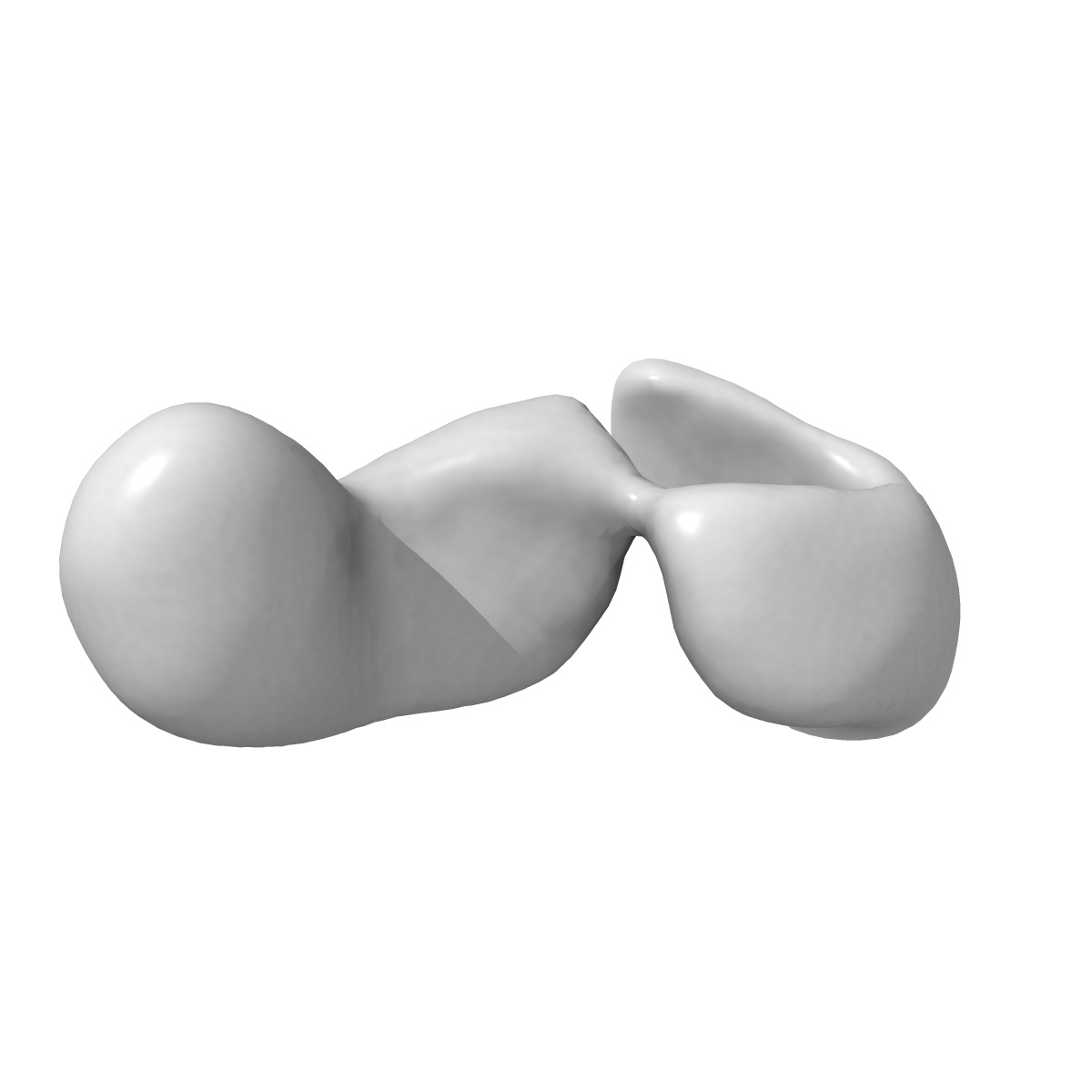
Negative stain electron microscopy structure of Nup192
EMD-5556
Single-particle26.0 Å
 Deposition: 17/12/2012
Map released: 26/12/2012
Last modified: 26/03/2014
Concentration: 0.0035
mg/mL
Buffer
pH: 7.4
Details: 20 mM HEPES, 300 mM NaCl, 2 mM MgCl2, 0.01% Tween 20, 0.01 mM DTT
Details: 20 mM HEPES, 300 mM NaCl, 2 mM MgCl2, 0.01% Tween 20, 0.01 mM DTT
Staining
Type:
NEGATIVE
Details: 3 ul protein was added to grids. The sample was blotted and 3 uL 1% uranyl formate added. Uranyl formate was blotted and fresh stain added (three times). The sample was allowed to sit 30 seconds before final blot, then air-dried.
Details: 3 ul protein was added to grids. The sample was blotted and 3 uL 1% uranyl formate added. Uranyl formate was blotted and fresh stain added (three times). The sample was allowed to sit 30 seconds before final blot, then air-dried.
Grid
Details: 200 mesh copper grid with thin carbon support, glow discharged 20s with Gatan Solarus 20s in H2/O2.
Microscope: JEOL 2100F
Illumination mode: FLOOD BEAM
Imaging mode: BRIGHT FIELD
Electron source: FIELD EMISSION GUN
Acceleration voltage: 200 kV
Nominal CS: 2 mm
Nominal defocus: 1.5 µm - 2.0 µm
Nominal magnification: 50000.0
Calibrated magnification: 81911.0
Specimen holder model: JEOL
Specimen holder details: High tilt
Alignment procedure: LEGACY (Astigmatism: Objective lens astigmatism was corrected at 250,000 times magnification, Electron beam tilt params: )
Minimum tilt angle: 0
Maximum tilt angle: 50
Details: Collected on 2028x2048 CCD, 24 um/pixel
Image Recording
[1]
Detector category:
CCD
Detector model: TVIPS TEMCAM-F224 (2k x 2k)
Sampling interval: 24 µm
Number of real images: 400
Average electron dose per image: 20 e/Å2
Bits per pixel: 16.0
Detector model: TVIPS TEMCAM-F224 (2k x 2k)
Sampling interval: 24 µm
Number of real images: 400
Average electron dose per image: 20 e/Å2
Bits per pixel: 16.0
Microscope: JEOL 2100F
Illumination mode: FLOOD BEAM
Imaging mode: BRIGHT FIELD
Electron source: FIELD EMISSION GUN
Acceleration voltage: 200 kV
Nominal CS: 2 mm
Nominal defocus: 1.5 µm - 2.0 µm
Nominal magnification: 50000.0
Calibrated magnification: 81911.0
Specimen holder model: JEOL
Specimen holder details: High tilt
Alignment procedure: LEGACY (Astigmatism: Objective lens astigmatism was corrected at 250,000 times magnification, Electron beam tilt params: )
Minimum tilt angle: 0
Maximum tilt angle: 50
Details: Collected on 2028x2048 CCD, 24 um/pixel. 2 sessions are listed; data collected on multiple days between these dates
Image Recording
[1]
Detector category:
CCD
Detector model: TVIPS TEMCAM-F224 (2k x 2k)
Sampling interval: 24 µm
Number of real images: 400
Average electron dose per image: 20 e/Å2
Bits per pixel: 16.0
Detector model: TVIPS TEMCAM-F224 (2k x 2k)
Sampling interval: 24 µm
Number of real images: 400
Average electron dose per image: 20 e/Å2
Bits per pixel: 16.0
Details: Particles were selected manually using jweb and boxer
Final
reconstruction
Resolution: 26.0
Å
(
BY AUTHOR)
Resolution method: FSC 0.5 CUT-OFF
Number of images used: 3883
Algorithm: OTHER
Details: Initial map was calculated by merging 3 maps obtained from random conical tilt. This was then used for 8 rounds of reference-based refinement in SPIDER at a 10-degree angular increment
Resolution method: FSC 0.5 CUT-OFF
Number of images used: 3883
Algorithm: OTHER
Details: Initial map was calculated by merging 3 maps obtained from random conical tilt. This was then used for 8 rounds of reference-based refinement in SPIDER at a 10-degree angular increment
⌯ Applied Symmetry
Point group:
C1
Software
[1]
| Name | Version | Details |
|---|---|---|
| Spider, Sparx, EMAN2, EMAN | - | - |
⦩ Final angle assignment
Details: SPIDER: theta 45 degrees, phi 45 degrees
Final 2D classification
Number of classes:
23
CTF correction
Details:Phase flip each particle
Format: CCP4
Data type: IMAGE STORED AS FLOATING POINT NUMBER (4 BYTES)
Annotation details: Reconstruction of full-length Nup192
Details: ::::EMDATABANK.org::::EMD-5556::::
Data type: IMAGE STORED AS FLOATING POINT NUMBER (4 BYTES)
Annotation details: Reconstruction of full-length Nup192
Details: ::::EMDATABANK.org::::EMD-5556::::
⬡ Geometry
| X | Y | Z | |
|---|---|---|---|
| Dimensions | 80 | 80 | 80 |
| Origin | 0 | 0 | 0 |
| Spacing | 80 | 80 | 80 |
| Voxel size | 2.93 Å | 2.93 Å | 2.93 Å |
Contour list
| Primary | Level | Source |
|---|---|---|
| True | 0.0721 | AUTHOR |