EMD-5590
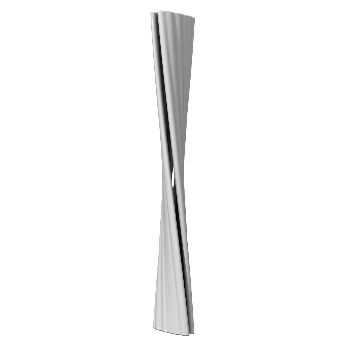
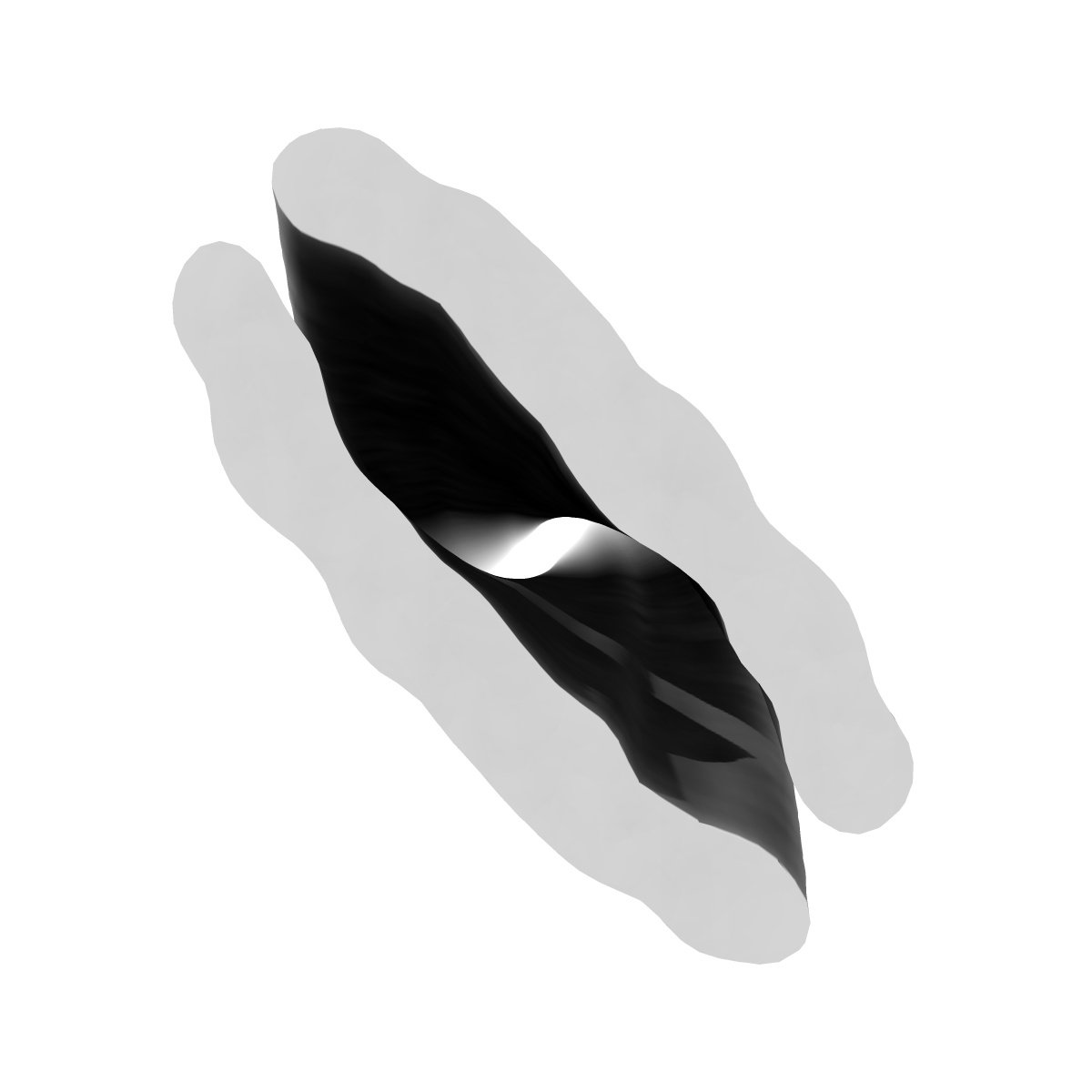
Electron cryo-microscopy of a cross-beta amyloid fibril polymorph
EMD-5590
Single-particle12.7 Å
 Deposition: 27/02/2013
Map released: 03/04/2013
Last modified: 06/11/2013
Concentration: 1
mg/mL
Buffer
pH: 2.0
Details: 10% acetonitrile/water solution
Details: 10% acetonitrile/water solution
Grid
Details: Holey carbon films (R2/2, QuantifoilMicro Tools GmbH, Jena, Germany)
Vitrification
Cryogen name: ETHANE
Chamber humidity: 95%
Chamber temperature: 99 K
Instrument: HOMEMADE PLUNGER
Method: Fibrils were applied to holey carbon films that were immediately plunge-frozen at liquid nitrogen temperature.
Chamber humidity: 95%
Chamber temperature: 99 K
Instrument: HOMEMADE PLUNGER
Method: Fibrils were applied to holey carbon films that were immediately plunge-frozen at liquid nitrogen temperature.
Microscope: FEI TECNAI F20
Illumination mode: FLOOD BEAM
Imaging mode: BRIGHT FIELD
Electron source: FIELD EMISSION GUN
Acceleration voltage: 200 kV
Nominal defocus: 0.9 µm - 3.0 µm
Nominal magnification: 40000.0
Calibrated magnification: 40000.0
Specimen holder model: GATAN LIQUID NITROGEN
Specimen holder details: Nitrogen cooled
Alignment procedure: LEGACY (Astigmatism: Objective lens astigmatism was corrected at 100,000 times magnification., Electron beam tilt params: )
Minimum tilt angle: 0
Maximum tilt angle: 0
Temperature
Minimum: 99
K
Average: 99 K
Maximum: 100 K
Average: 99 K
Maximum: 100 K
Image Recording
[1]
Detector category:
FILM
Detector model: KODAK SO-163 FILM
Scanner: ZEISS SCAI
Number of real images: 250
Detector model: KODAK SO-163 FILM
Scanner: ZEISS SCAI
Number of real images: 250
Details: The particles were selected using an automatic selection program.
Final
reconstruction
Resolution: 12.7
Å
(
BY AUTHOR)
Resolution method: FSC 0.5 CUT-OFF
Number of images used: 82
Algorithm: OTHER
Details:
Resolution method: FSC 0.5 CUT-OFF
Number of images used: 82
Algorithm: OTHER
Details:
⌯ Applied Symmetry
ΔΖ:
4.67
Å
ΔΦ: 0.85°
ΔΦ: 0.85°
Software
[1]
| Name | Version | Details |
|---|---|---|
| SPIDER, IMAGIC, BKRP | - | - |
Final 2D classification
Number of classes:
18
CTF correction
Details:CTFFIND3
Format: CCP4
Data type: IMAGE STORED AS FLOATING POINT NUMBER (4 BYTES)
Annotation details: Reconstruction of doublet cross-beta amyloid fibril polymorph
Details: ::::EMDATABANK.org::::EMD-5590::::
Data type: IMAGE STORED AS FLOATING POINT NUMBER (4 BYTES)
Annotation details: Reconstruction of doublet cross-beta amyloid fibril polymorph
Details: ::::EMDATABANK.org::::EMD-5590::::
⬡ Geometry
| X | Y | Z | |
|---|---|---|---|
| Dimensions | 120 | 120 | 298 |
| Origin | -60 | -59 | -149 |
| Spacing | 120 | 120 | 298 |
| Voxel size | 1.8 Å | 1.8 Å | 1.7999998 Å |
Contour list
| Primary | Level | Source |
|---|---|---|
| True | 230.43 | AUTHOR |