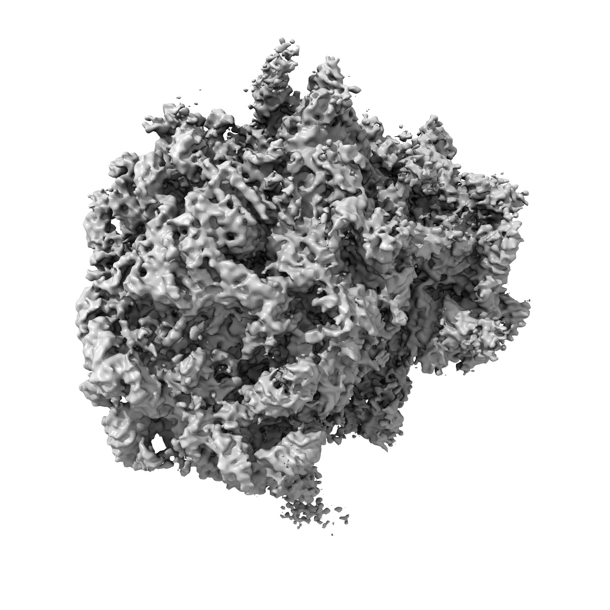
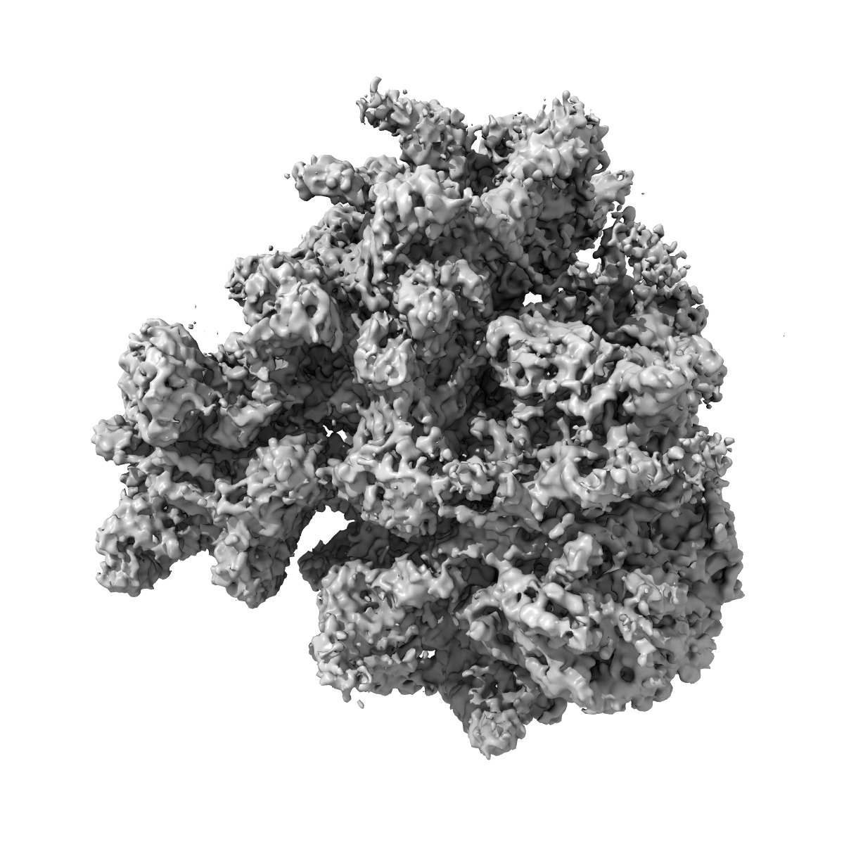
EMD-5591
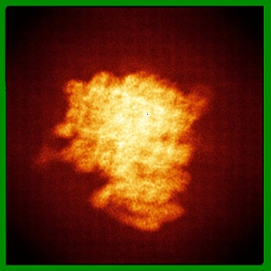
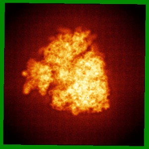
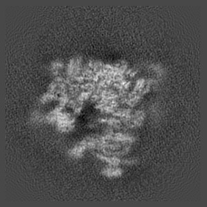
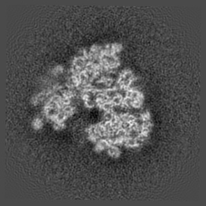
Electron cryo-microscopy of Drosophila melanogaster EF2- and Vig2-bound 80S ribosome
EMD-5591
Single-particle6.0 Å
 Deposition: 28/02/2013
Map released: 01/05/2013
Last modified: 23/07/2014
Buffer
pH: 7.5
Vitrification
Cryogen name: ETHANE
Chamber humidity: 100%
Instrument: FEI VITROBOT MARK IV
Method: Before plunging, blot for 3 seconds using two layers of filter paper.
Chamber humidity: 100%
Instrument: FEI VITROBOT MARK IV
Method: Before plunging, blot for 3 seconds using two layers of filter paper.
Microscope: FEI TITAN KRIOS
Illumination mode: FLOOD BEAM
Imaging mode: BRIGHT FIELD
Electron source: FIELD EMISSION GUN
Acceleration voltage: 300 kV
Nominal CS: 2.7 mm
Nominal defocus: 1.0 µm - 4.0 µm
Nominal magnification: 90000.0
Calibrated magnification: 90000.0
Specimen holder model: GATAN LIQUID NITROGEN
Illumination mode: FLOOD BEAM
Imaging mode: BRIGHT FIELD
Electron source: FIELD EMISSION GUN
Acceleration voltage: 300 kV
Nominal CS: 2.7 mm
Nominal defocus: 1.0 µm - 4.0 µm
Nominal magnification: 90000.0
Calibrated magnification: 90000.0
Specimen holder model: GATAN LIQUID NITROGEN
Image Recording
[1]
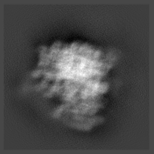
Details: The particles were selected using an automatic selection program
Final
reconstruction
Resolution: 6.0
Å
(
BY AUTHOR)
Resolution method: FSC 0.5 CUT-OFF
Number of images used: 134500
Algorithm: OTHER
Details:
Resolution method: FSC 0.5 CUT-OFF
Number of images used: 134500
Algorithm: OTHER
Details:
⌯ Applied Symmetry
Point group:
C1
Software
[1]
| Name | Version | Details |
|---|---|---|
| Spider | - | - |
CTF correction
Details:Each subvolume
Format: CCP4
Data type: IMAGE STORED AS FLOATING POINT NUMBER (4 BYTES)
Annotation details: Reconstruction of D. melanogaster 80S ribosome
Details: ::::EMDATABANK.org::::EMD-5591::::
Data type: IMAGE STORED AS FLOATING POINT NUMBER (4 BYTES)
Annotation details: Reconstruction of D. melanogaster 80S ribosome
Details: ::::EMDATABANK.org::::EMD-5591::::
⬡ Geometry
| X | Y | Z | |
|---|---|---|---|
| Dimensions | 368 | 368 | 368 |
| Origin | -184 | -184 | -183 |
| Spacing | 368 | 368 | 368 |
| Voxel size | 1.2375 Å | 1.2375 Å | 1.2375 Å |
Contour list
| Primary | Level | Source |
|---|---|---|
| True | 0.49 | AUTHOR |