EMD-5601
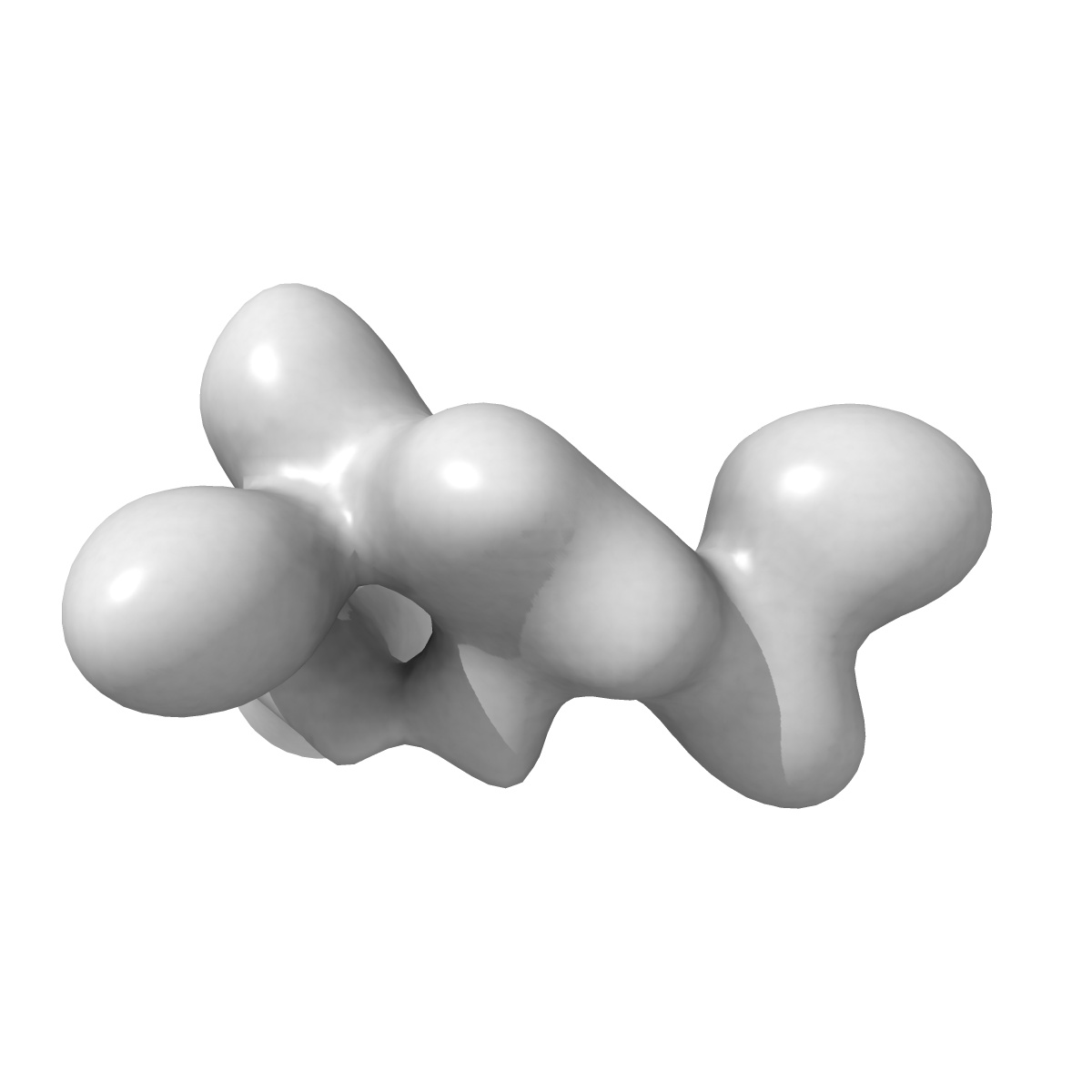
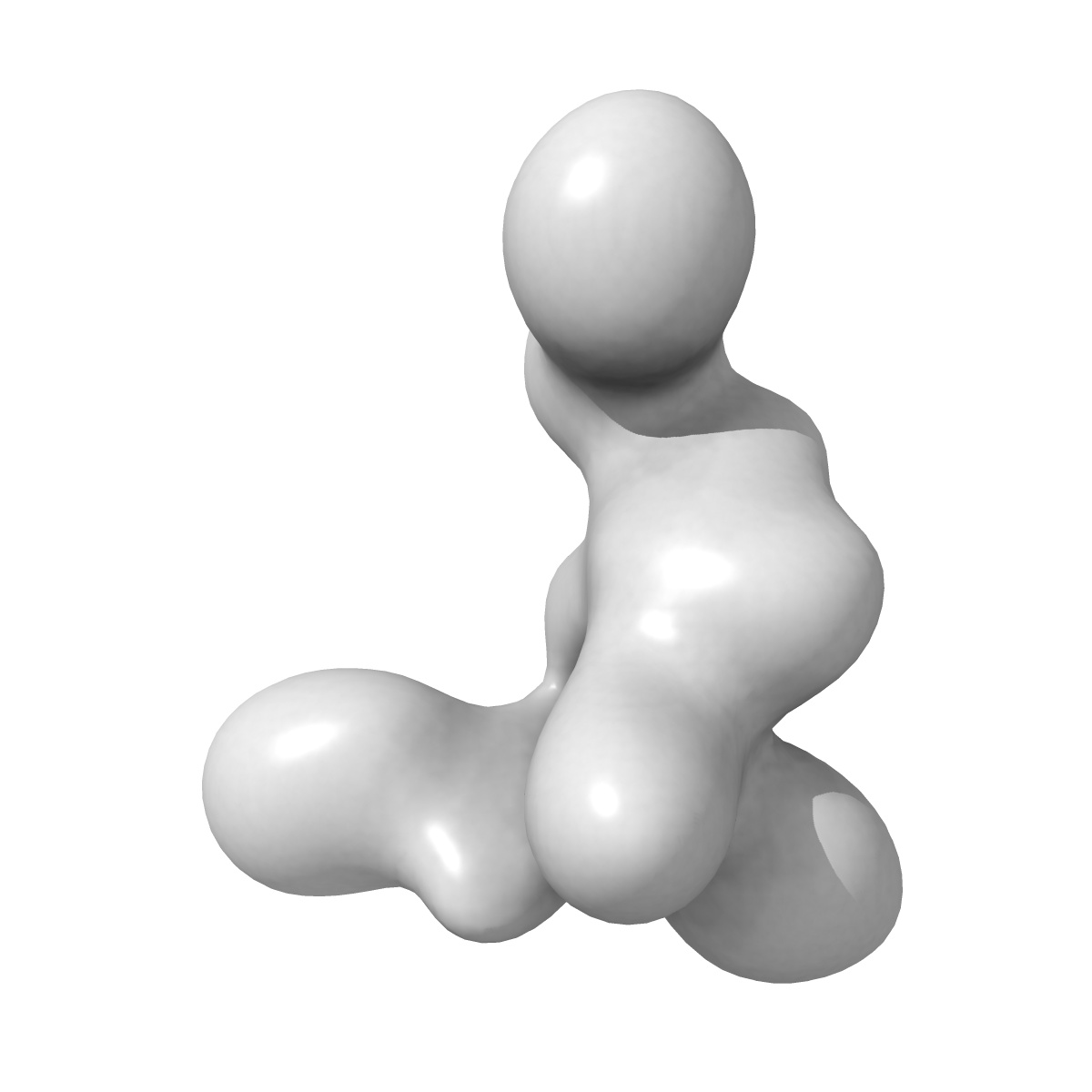
Substrate-specific structural rearrangements of human Dicer
EMD-5601
Single-particle26.0 Å
 Deposition: 09/03/2013
Map released: 01/05/2013
Last modified: 19/06/2013
Concentration: 0.05
mg/mL
Buffer
pH: 7.4
Details: 20 mM HEPES, pH 7.5, 150 mM KCl, 3 mM EDTA, 1 mM DTT, and 2.5% glycerol
Details: 20 mM HEPES, pH 7.5, 150 mM KCl, 3 mM EDTA, 1 mM DTT, and 2.5% glycerol
Grid
Details: Glow-discharged Quantifoil R 1.2/1.3 MO 200 mesh holey carbon grids
Vitrification
Cryogen name: ETHANE
Chamber humidity: 100%
Instrument: FEI VITROBOT MARK IV
Method: The samples were automatically blotted for 4-5 s at -2.5 mm offset before plunging.
Chamber humidity: 100%
Instrument: FEI VITROBOT MARK IV
Method: The samples were automatically blotted for 4-5 s at -2.5 mm offset before plunging.
Microscope: JEOL 3100FFC
Illumination mode: FLOOD BEAM
Imaging mode: BRIGHT FIELD
Electron source: FIELD EMISSION GUN
Acceleration voltage: 300 kV
Nominal CS: 3.7 mm
Nominal defocus: 0.0 µm - 0.0 µm
Nominal magnification: 60000.0
Calibrated magnification: 100000.0
Specimen holder model: JEOL
Specimen holder details: Liquid helium cooled stage maintained at 55 K
Alignment procedure: LEGACY (Astigmatism: Objective lens astigmatism was corrected at 100,000 times magnification, Electron beam tilt params: )
Temperature
Minimum: 45
K
Average: 55 K
Maximum: 60 K
Average: 55 K
Maximum: 60 K
Specialist optics
Energy filter
Image Recording
[1]
Detector category:
CCD
Detector model: GENERIC GATAN (2k x 2k)
Number of real images: 800
Average electron dose per image: 20 e/Å2
Detector model: GENERIC GATAN (2k x 2k)
Number of real images: 800
Average electron dose per image: 20 e/Å2
Final
reconstruction
Resolution: 26.0
Å
(
BY AUTHOR)
Resolution method: FSC 0.5 CUT-OFF
Number of images used: 4800
Algorithm: OTHER
Resolution method: FSC 0.5 CUT-OFF
Number of images used: 4800
Algorithm: OTHER
⌯ Applied Symmetry
Point group:
C1
Software
[1]
| Name | Version | Details |
|---|---|---|
| SPIDER | - | - |
CTF correction
Details:Each micrograph
Format: CCP4
Data type: IMAGE STORED AS FLOATING POINT NUMBER (4 BYTES)
Annotation details: Zernike phase-constrast cryo-EM reconstruction of human Dicer
Details: ::::EMDATABANK.org::::EMD-5601::::
Data type: IMAGE STORED AS FLOATING POINT NUMBER (4 BYTES)
Annotation details: Zernike phase-constrast cryo-EM reconstruction of human Dicer
Details: ::::EMDATABANK.org::::EMD-5601::::
⬡ Geometry
| X | Y | Z | |
|---|---|---|---|
| Dimensions | 90 | 90 | 90 |
| Origin | -45 | -45 | -45 |
| Spacing | 90 | 90 | 90 |
| Voxel size | 3.07 Å | 3.07 Å | 3.07 Å |
Contour list
| Primary | Level | Source |
|---|---|---|
| True | 2.07 | AUTHOR |