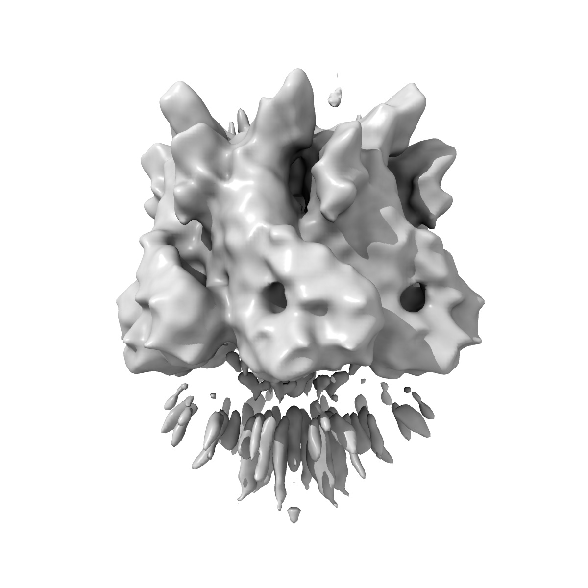
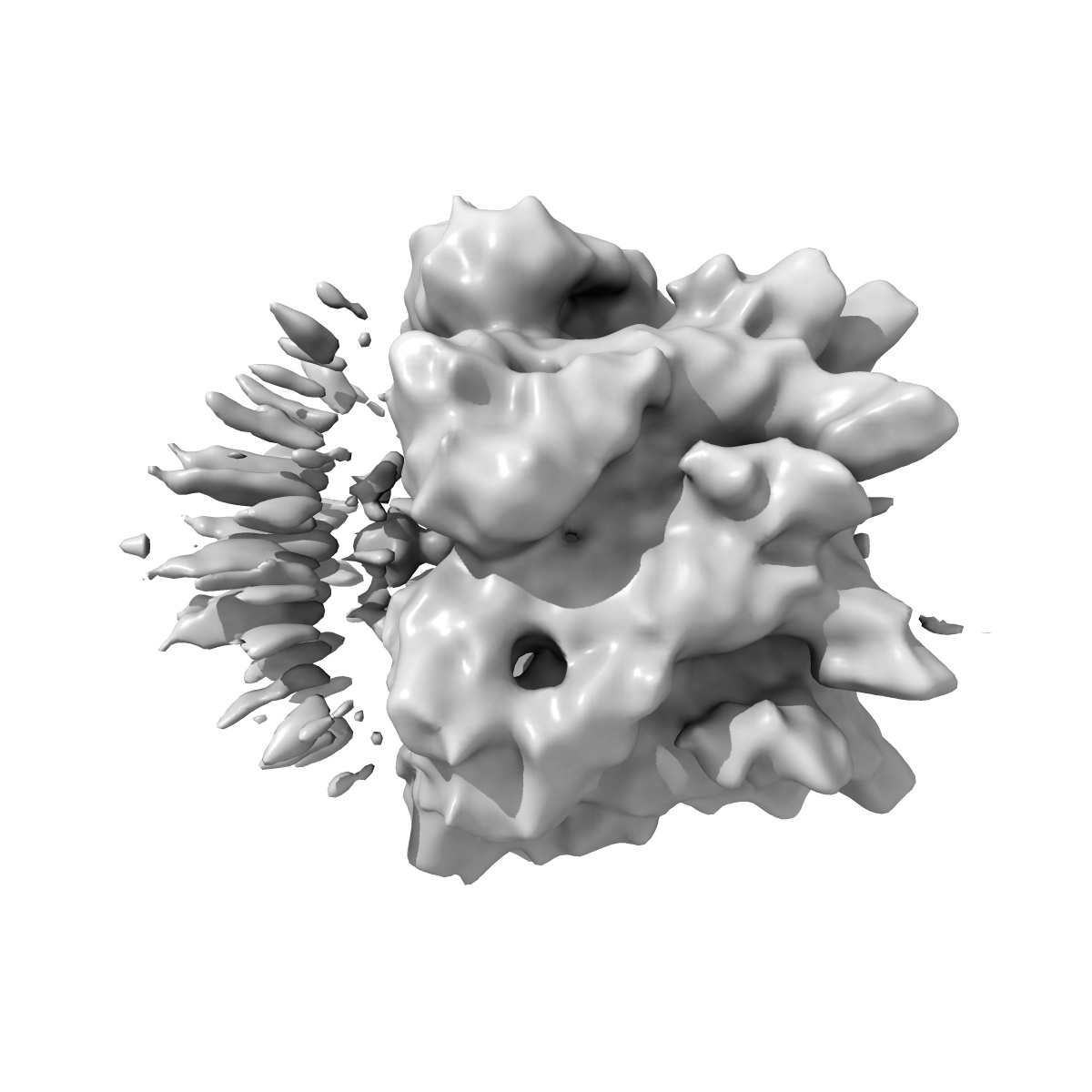
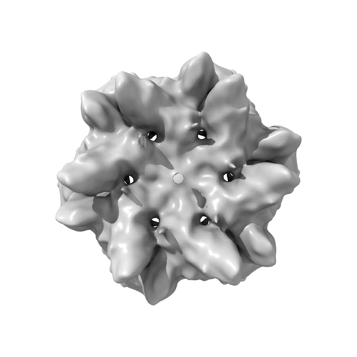
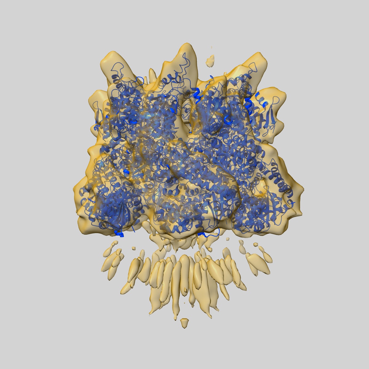
EMD-5607
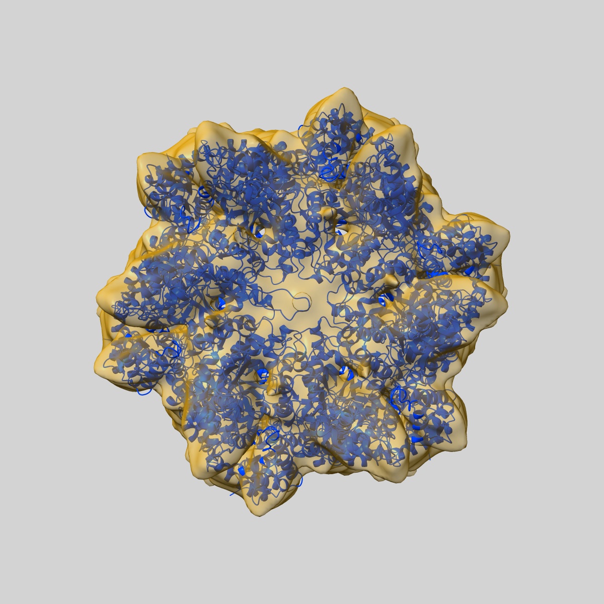
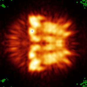
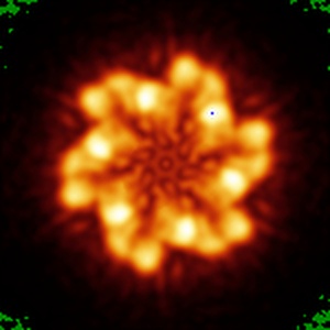
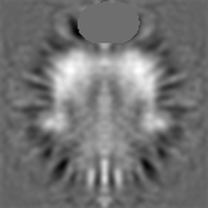
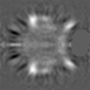
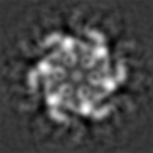
Structural dynamics and inter-ring communication of the MecA-ClpC complex during active substrate unfolding and translocation revealed by cryo-EM
EMD-5607
Single-particle9.0 Å
 Deposition: 20/03/2013
Map released: 15/05/2013
Last modified: 28/08/2013
Concentration: 0.03
mg/mL
Buffer
pH: 7.5
Details: 50mM kCl, 10mM Tris-HCL,2mM MgCl2, 2mM ATP
Details: 50mM kCl, 10mM Tris-HCL,2mM MgCl2, 2mM ATP
Vitrification
Cryogen name: ETHANE
Chamber humidity: 100%
Chamber temperature: 90 K
Instrument: FEI VITROBOT MARK IV
Method: Blot for 2 seconds before plunging
Chamber humidity: 100%
Chamber temperature: 90 K
Instrument: FEI VITROBOT MARK IV
Method: Blot for 2 seconds before plunging
Microscope: FEI TITAN KRIOS
Illumination mode: FLOOD BEAM
Imaging mode: BRIGHT FIELD
Electron source: FIELD EMISSION GUN
Acceleration voltage: 300 kV
Nominal defocus: 1.5 µm - 3.0 µm
Nominal magnification: 59000.0
Specimen holder model: FEI TITAN KRIOS AUTOGRID HOLDER
Illumination mode: FLOOD BEAM
Imaging mode: BRIGHT FIELD
Electron source: FIELD EMISSION GUN
Acceleration voltage: 300 kV
Nominal defocus: 1.5 µm - 3.0 µm
Nominal magnification: 59000.0
Specimen holder model: FEI TITAN KRIOS AUTOGRID HOLDER
Image Recording
[1]
Detector category:
CCD
Detector model: FEI EAGLE (4k x 4k)
Number of real images: 994
Average electron dose per image: 20 e/Å2
Detector model: FEI EAGLE (4k x 4k)
Number of real images: 994
Average electron dose per image: 20 e/Å2
Final
reconstruction
Resolution: 9.0
Å
(
BY AUTHOR)
Resolution method: FSC 0.5 CUT-OFF
Number of images used: 45514
Algorithm: OTHER
Resolution method: FSC 0.5 CUT-OFF
Number of images used: 45514
Algorithm: OTHER
⌯ Applied Symmetry
Point group:
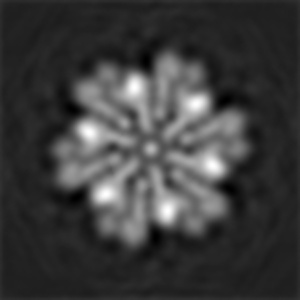
C6
Software
[1]
| Name | Version | Details |
|---|---|---|
| SPIDER | - | - |
CTF correction
Details:Each defocus group on 3D level
Format: CCP4
Data type: IMAGE STORED AS FLOATING POINT NUMBER (4 BYTES)
Annotation details: Reconstruction of MecA-ClpC(E280A) with ATP, MW-ATP
Details: ::::EMDATABANK.org::::EMD-5607::::
Data type: IMAGE STORED AS FLOATING POINT NUMBER (4 BYTES)
Annotation details: Reconstruction of MecA-ClpC(E280A) with ATP, MW-ATP
Details: ::::EMDATABANK.org::::EMD-5607::::
⬡ Geometry
| X | Y | Z | |
|---|---|---|---|
| Dimensions | 150 | 150 | 150 |
| Origin | -74 | -74 | -74 |
| Spacing | 150 | 150 | 150 |
| Voxel size | 1.5 Å | 1.5 Å | 1.5 Å |
Contour list
| Primary | Level | Source |
|---|---|---|
| True | 1.5 | AUTHOR |