EMD-5743
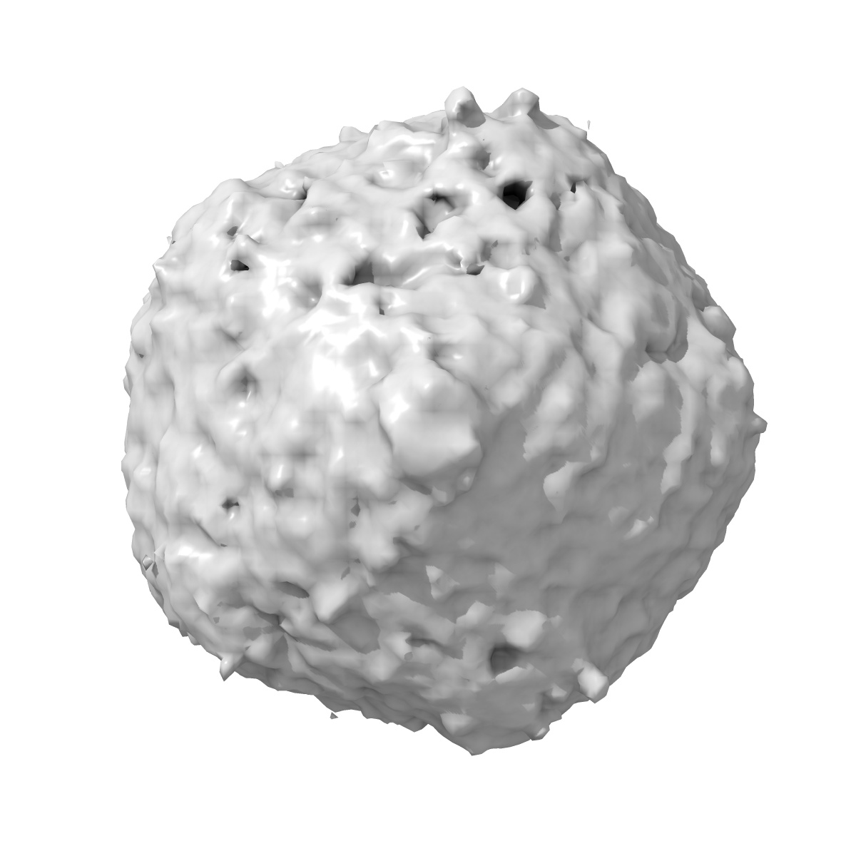
Zernike Phase Contrast electron cryo-tomography of cyanophage Syn5 assembly intermediates
EMD-5743
Subtomogram averaging74.0 Å
 Deposition: 13/08/2013
Map released: 09/10/2013
Last modified: 06/11/2013
Buffer
pH: 8.0
Details: Artificial sea water
Details: Artificial sea water
Grid
Details: 200 mesh Quantifoil holey grids, glow discharged
Vitrification
Cryogen name: ETHANE
Chamber humidity: 95%
Chamber temperature: 85 K
Instrument: FEI VITROBOT MARK III
Method: Blot for 3 seconds before plunging
Chamber humidity: 95%
Chamber temperature: 85 K
Instrument: FEI VITROBOT MARK III
Method: Blot for 3 seconds before plunging
Microscope: JEOL 2200FS
Illumination mode: FLOOD BEAM
Imaging mode: BRIGHT FIELD
Electron source: FIELD EMISSION GUN
Acceleration voltage: 200 kV
Nominal CS: 4.1 mm
Nominal defocus: 0.0 µm - 1.0 µm
Nominal magnification: 25000.0
Calibrated magnification: 25000.0
Specimen holder model: GATAN LIQUID NITROGEN
Alignment procedure: LEGACY (Astigmatism: Objective lens astigmatism was corrected at 100,000 times magnification, Electron beam tilt params: )
Temperature
Minimum: 93
K
Average: 103 K
Maximum: 105 K
Average: 103 K
Maximum: 105 K
Specialist optics
Energy filter
Image Recording
[1]
Detector category:
CCD
Detector model: GATAN ULTRASCAN 4000 (4k x 4k)
Number of real images: 42
Average electron dose per image: 1 e/Å2
Detector distance: 1200
Bits per pixel: 16.0
Detector model: GATAN ULTRASCAN 4000 (4k x 4k)
Number of real images: 42
Average electron dose per image: 1 e/Å2
Detector distance: 1200
Bits per pixel: 16.0
Tilt Series
[1]
| Axis 1 | Axis 2 | |||||
|---|---|---|---|---|---|---|
| Min. | Max. | Inc. | Min. | Max. | Inc. | Rotation |
| -60 ° | 60 ° | - | - | - | - | - |
Details: The subtomograms were extracted from infected cells.
Final
reconstruction
Resolution: 74.0
Å
(
BY AUTHOR)
Resolution method: FSC 0.143 CUT-OFF
Algorithm: OTHER
Details:
Resolution method: FSC 0.143 CUT-OFF
Algorithm: OTHER
Details:
⌯ Applied Symmetry
Point group:
C1
Software
[1]
| Name | Version | Details |
|---|---|---|
| IMOD | - | - |
Final 3D classification
Number of classes:
1
Format: CCP4
Data type: IMAGE STORED AS FLOATING POINT NUMBER (4 BYTES)
Annotation details: Cyanophage Syn5 assembly intermediate - the expanded capsid
Details: ::::EMDATABANK.org::::EMD-5743::::
Data type: IMAGE STORED AS FLOATING POINT NUMBER (4 BYTES)
Annotation details: Cyanophage Syn5 assembly intermediate - the expanded capsid
Details: ::::EMDATABANK.org::::EMD-5743::::
⬡ Geometry
| X | Y | Z | |
|---|---|---|---|
| Dimensions | 128 | 128 | 128 |
| Origin | 32 | 32 | 32 |
| Spacing | 128 | 128 | 128 |
| Voxel size | 9.04 Å | 9.04 Å | 9.04 Å |
Contour list
| Primary | Level | Source |
|---|---|---|
| True | 1.9 | AUTHOR |