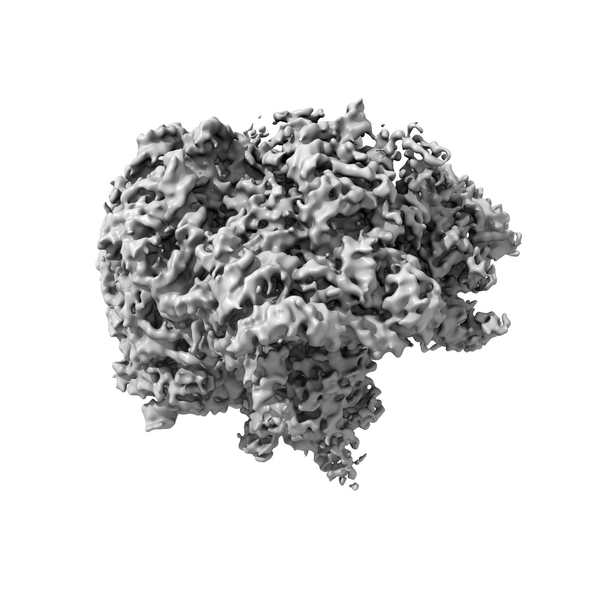
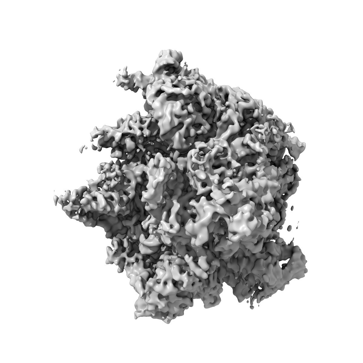
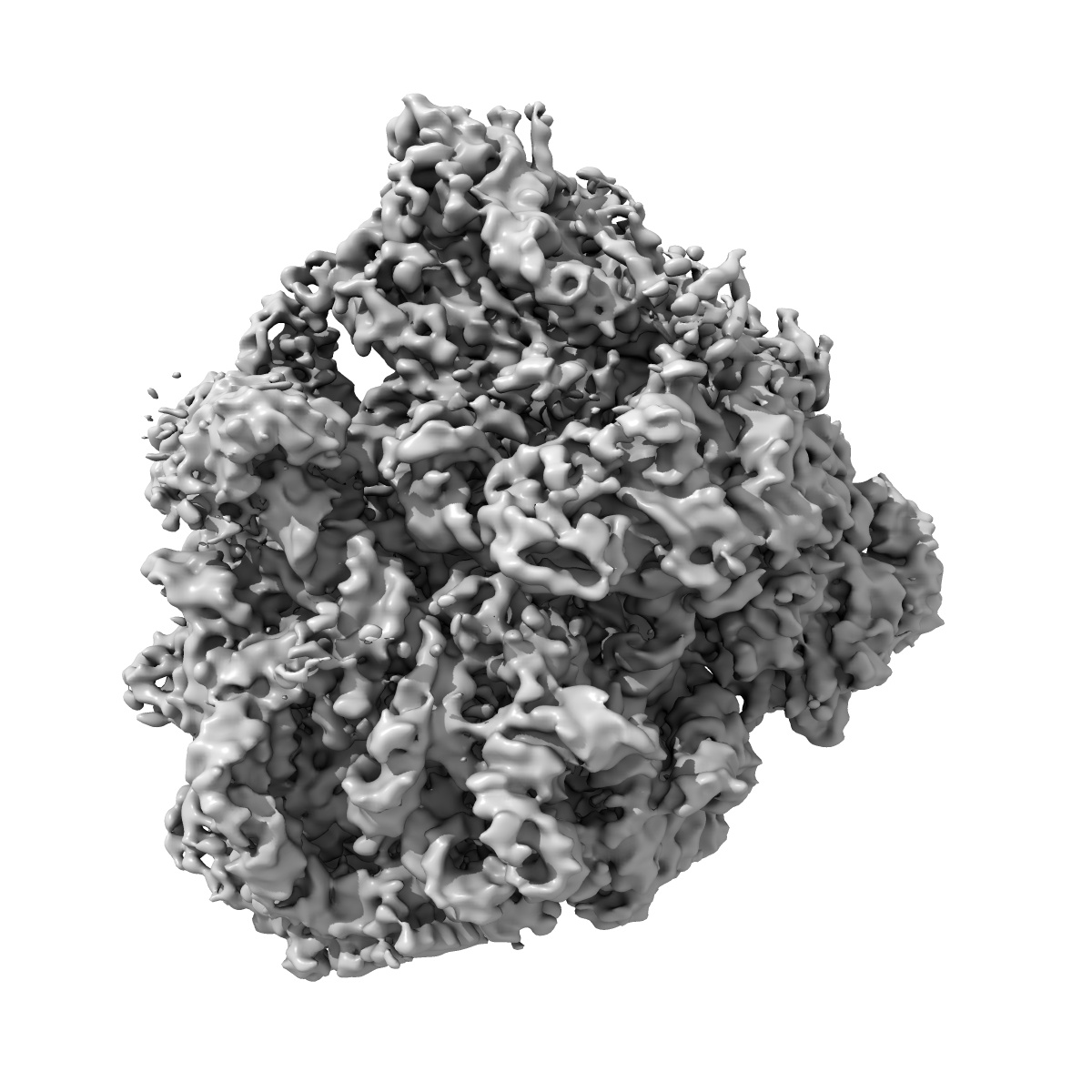
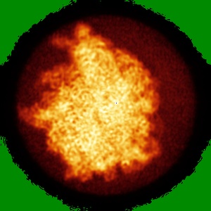
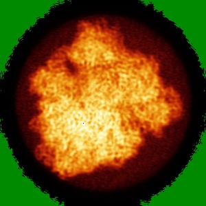
EMD-5797
Structure of the Ribosome with Elongation Factor G Trapped in the Pre-Translocation State
EMD-5797
Single-particle6.2 Å
 Deposition: 19/11/2013
Map released: 18/12/2013
Last modified: 15/01/2014
Concentration: 0.4
mg/mL
Buffer
pH: 7.6
Details: 10 mM HEPES-KOH, 5 mM MgCl2, 90 mM NH4Cl, 2 mM spermidine, 0.1 mM spermine, 6 mM BME, 0.5 mM viomycin, 0.5 mM GTP, 0.5 mM fusidic acid
Details: 10 mM HEPES-KOH, 5 mM MgCl2, 90 mM NH4Cl, 2 mM spermidine, 0.1 mM spermine, 6 mM BME, 0.5 mM viomycin, 0.5 mM GTP, 0.5 mM fusidic acid
Grid
Details: C-flat 1.2/1.3 holey carbon 400 mesh copper grid, glow discharged with a current of -20 mA for 45 seconds in an EMITECH K100X glow discharge unit
Vitrification
Cryogen name: ETHANE
Chamber humidity: 95%
Instrument: FEI VITROBOT MARK II
Method: Freshly glow-disharged grids were loaded into an FEI Mark II Vitrobot and equilibrated to 95% relative humidity at 22 degrees Celsius. 2 microliters of sample was applied through the side port, blotted for 7 seconds with a positional offset of 2, and plunged into liquid ethane.
Microscope: FEI TITAN KRIOS
Illumination mode: FLOOD BEAM
Imaging mode: BRIGHT FIELD
Electron source: FIELD EMISSION GUN
Acceleration voltage: 300 kV
Nominal CS: 0.01 mm
Nominal defocus: 1.15 µm - 6.95 µm
Nominal magnification: 133333.0
Calibrated magnification: 134615.0
Specimen holder model: FEI TITAN KRIOS AUTOGRID HOLDER
Alignment procedure: LEGACY (Astigmatism: Automatically corrected using FEI software, Electron beam tilt params: )
Image Recording
[1]
Detector category:
CCD
Detector model: FEI FALCON I (4k x 4k)
Sampling interval: 14.0 µm
Number of real images: 13341
Average electron dose per image: 30 e/Å2
Bits per pixel: 16.0
Detector model: FEI FALCON I (4k x 4k)
Sampling interval: 14.0 µm
Number of real images: 13341
Average electron dose per image: 30 e/Å2
Bits per pixel: 16.0
Details: Refinement and 3D classification performed by Frealign. See primary citation Supplementary Information for details.
Final
reconstruction
Resolution: 6.2
Å
(
BY AUTHOR)
Resolution method: OTHER
Number of images used: 1341961
Algorithm: OTHER
Details: Refinement included data to 12 Angstrom resolution to limit FSC bias. See primary citation Supplementary Information for details.
Resolution method: OTHER
Number of images used: 1341961
Algorithm: OTHER
Details: Refinement included data to 12 Angstrom resolution to limit FSC bias. See primary citation Supplementary Information for details.
⌯ Applied Symmetry
Point group:
C1
Software
[1]
| Name | Version | Details |
|---|---|---|
| EMAN2, IMAGIC, FREALIGN, RSAMPLE, CTFFIND3 | - | - |
CTF correction
Details:CTFFIND3, FREALIGN per micrograph
Format: CCP4
Data type: IMAGE STORED AS FLOATING POINT NUMBER (4 BYTES)
Annotation details: Ribosome reconstruction
Details: ::::EMDATABANK.org::::EMD-5797::::
Data type: IMAGE STORED AS FLOATING POINT NUMBER (4 BYTES)
Annotation details: Ribosome reconstruction
Details: ::::EMDATABANK.org::::EMD-5797::::
⬡ Geometry
| X | Y | Z | |
|---|---|---|---|
| Dimensions | 320 | 320 | 320 |
| Origin | 0 | 0 | 0 |
| Spacing | 320 | 320 | 320 |
| Voxel size | 1.04 Å | 1.04 Å | 1.04 Å |
Contour list
| Primary | Level | Source |
|---|---|---|
| True | 0.4 | AUTHOR |