Documentation
Summary
- EMDB data model
- EMDB header data model
- EMDB segmentation data model
- Policies
- Search engine
- Chart builder
- FAQ
EMDB map data model
The EM Data Bank (EMDB) accepts and distributes 3D map volumes derived from several types of EM reconstruction methods, including single particle averaging, helical averaging, 2D crystallography, and tomography. Since its inception in 2002, the EMDB map distribution format has followed CCP4 definition (CCP4 map format) , which is widely recognized by software packages used by the structural biology community. CCP4 map format is closely related to the MRC map format used in the 3DEM community (MRC map format); CCP4 is slightly more restrictive, in that voxel positions are limited to a grid that includes the Cartesian coordinate origin (0,0,0). Further details can be found here.
EMDB header data model
Every EMDB entry has a header file containing meta data (e.g., sample, detector, microscope, image processing) describing the experiment. The header file is an XML file and the structure and content of the header file is described by a XSD data model. With a highly dynamic field such as cryo-EM there is a constant need to adapt and modify the schema to keep it up-to-date with the most recent developments. We consult extensively with the EM community regarding such issues and version the schema according to the policy described here.
Data model version 1.9
This has been a long-term stable version of the data model. It was be replaced in 2018 with an updated model but XML header files in version 1.9 continues to be distributed in parallel for at least one year to give EMDB users ample time to switch. It should be noted that the generation of the version 1.9 header files will be on a best effort basis but involves a back translation from recent versions that are richer in content and will therefore not contain all the information that can be found in the more recent versions.
Download schema
Browse schema documentation
Download Python code to facilitate reading and writing XML version 1.9 header files
Data model version 3.0 (current model)
This data model replaced version 1.9, however header files corresponding to both data models will be distributed in parallel with the view of stopping the distribution of the version 1.9 files in 2019 once users have had a chance to adopt version 3.0.
This version adds a number of features including:
- An improved description of direct electron detectors, specimen preparation and tomography experiments.
- A hierarchal description of the overall sample composition in combination with a low-level description of the macromolecular composition to allow the description of both molecular and cellular samples.
- Specific data items describing the half-maps and segmentations included with the entry.
Download schema
Browse schema documentation
Download Python code to facilitate reading and writing XML version 1.9 header files
EMDB segmentation data model
Segmentation is the decomposition of 3D volumes into regions that can be associated with defined objects. Following several consultations with the EM community (Patwardhan et al., 2012; Patwardhan et al., 2014; Patwardhan et al., 2017), the EMDB is in the process of developing tools to support deposition of volume segmentations with structured biological annotation which is here defined as the association of data with identifiers (e.g., accession codes from UniProt) and ontologies taken from well established bioinformatics resources. To our knowledge, none of the segmentation formats widely used in electron microscopy and related fields currently support structured biological annotation. Third party use of segmentations is further impeded by the prevalence of segmentation file formats and their lack of interoperability. EMDB therefore proposed an open segmentation file format called EMDB-SFF to capture basic segmentation data from application-specific segmentation file formats and provide the means for structured biological annotation. In this way, EMDB-SFF will not only enable depositions of segmentations but also act as a file interchange format between different applications and facilitate analysis of 3D reconstructions. Furthermore EMDB-SFF supports the description of multiple transforms for a segment, thus allowing a segment to be used to describe the placement of a sub-tomogram average onto a tomographic reconstruction.
Model
EMDB-SFF files have the follow features:
- Segmentation metadata:
- name
- version (of schema)
- details (free-form text)
- global external references, e.g. specimen scientific identifier
- bounding box
- primary descriptor contained i.e. one of ‘three_d_volume’, ‘mesh_list’, or ‘shape_primitive_list’ (see schema documentation)
- list of software used to create the segmentation (name, version, processing details)
- list of transforms referenced by segments e.g. transform to place the sub-tomogram average in the tomogram
- Hierarchical ordering of segments through the use of segment IDs and parent IDs;
- Four geometrical representations of segments (volumes, contours, meshes, shapes);
- Can store subtomogram averages and how they map into the parent tomogram through the use of transforms;
- List of associated external references per segment;
- List of associated complexes and macromolecules in a related EMDB entry
Each segment in a segmentation can consist of two types of descriptors:
- textual descriptors;
- geometric descriptors.
Textual descriptors consist of either free-form text or standardised terms. Standard terms should be provided from a [published] ontology or list of identifiers.
Geometric descriptors can take one or more of the following representations:
- ‘three_d_volume’ for 3D volumes;
- ‘mesh_list’ for lists of meshes each of which consists of a set of vertices and polygons;
- lists of shape primitives (ellipsoid, cuboid, cone, cylinder).
Documentation
Download
The current schema (version 0.8.0.dev1) is available here.
Documentation
Complete documentation of the schema is available here.
Auxiliary Tools
sfftk-rw
sfftk-rw is a Python toolkit for reading and writing EMDB-SFF files only. It is part of a family of tools designed to work with EMDB-SFF files.
sfftk-rw has the following utilities:
- convert - interconvert between XML, HDF5 and JSON file formats of the EMDB-SFF data model;
- view - view a file summary
The full documentation is available at readthedocs.
Download
The latest version runs only on Python 3 (version 0.7.1) and may be installed using pip install sfftk-rw. Alternatively, feel free to obtain the source code from Github.
sfftk
sfftk provides a shell command and a Python API to process EMDB-SFF files.
The following utilities are available using sfftk:
- convert - Conversion of application-specific segmentation file formats to EMDB-SFF. Currently, sfftk supports the following formats:
- AmiraMesh (.am)
- Amira HyperSurface (.surf)
- Segger (.seg)
- EMDB Map masks (.map)
- Stereolithography (.stl)
- IMOD (.mod)
- notes - Annotation of EMDB-SFF files.
- view - Brief summaries of segmentation files.
Read the full documentation here.
Download
The latest development version (version 0.5.5.dev1) of sfftk may be downloaded/installed from PyPI or the source may be obtained from GitHub.
Publications
- Patwardhan, Ardan, Robert Brandt, Sarah J. Butcher, Lucy Collinson, David Gault, Kay Grünewald, Corey Hecksel et al. Building bridges between cellular and molecular structural biology. eLife 6 (2017).
- Patwardhan, Ardan, Alun Ashton, Robert Brandt, Sarah Butcher, Raffaella Carzaniga, Wah Chiu, Lucy Collinson et al. A 3D cellular context for the macromolecular world. Nature structural & molecular biology 21, no. 10 (2014): 841-845.
- Patwardhan, Ardan, José-Maria Carazo, Bridget Carragher, Richard Henderson, J. Bernard Heymann, Emma Hill, Grant J. Jensen et al. Data management challenges in three-dimensional EM. Nature structural & molecular biology 19, no. 12 (2012): 1203-1207.
Quick links
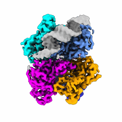
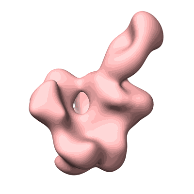
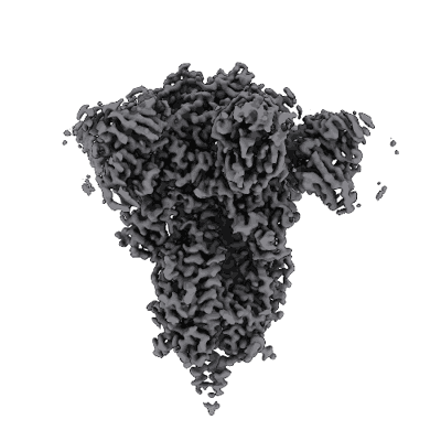
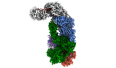
Recent Entries
(Show all)
Human 3-methylcrotonyl-CoA carboxylase in BCCP-CTS state with substrate

Consensus map: CW Flagellar Switch Complex - FliF, FliG, FliM, and FliN forming the C-ring from Salmonella


Cryo-EM structure of human high-voltage activated L-type calcium channel CaV1.2 (apo)
CRISPR-Cas type III-D effector complex bound to self-target RNA in a post-cleavage state

Influenza virus neuraminidase N1 NC13 ectodomain with a tetrabrachio-domain stalk
Low-dose cryo-electron ptychographic reconstruction of TMV recorded with CSA of 6.1 mrad

Cryo-EM structure of synthetic claudin-4 complex with Clostridium perfringens enterotoxin C-terminal domain, sFab COP-2, and Nanobody

Human parainfluenza virus type 3 prefusion F trimer in complex with rPIV3-18 Fab


Cryo-EM structure of human high-voltage activated L-type calcium channel CaV1.2 in complex with benidipine (BEN)

Structure of a 1033 Scaffold Base DNA Origami Nanostructure V4 with HPLC Purified Staples

Structure of the SFTSV L protein in a transcription-priming state with bound capped RNA [TRANSCRIPTION-PRIMING]

in-situ subtomogram average of C. elegans centrioles in centrosomes


Structure of Circularly Permuted 50S Ribosomal Subunit Assembly Intermediate - CP45 Class B

polysome/di-ribosome class I in chloramphenicol-treated Mycoplasma pneumoniae cells

Structure of human gamma-secretase PSEN1 APH-1B isoform reconstituted into lipid nanodisc in complex with Ab46
Cryo-EM structure of pre-subcore from in vitro assembled particles


70S ribosome with A and P-site tRNAs in chloramphenicol-treated Mycoplasma pneumoniae cells, K3 data

Structure of Circularly Permuted 50S Ribosomal Subunit Assembly Intermediate - CP63 Class C


CryoEM map of a de novo designed T=4 icosahedral nanocage hierarchically built from pseudosymmetric trimers; design Ico(T=4)-4

CRISPR-Cas type III-D effector complex bound to a target RNA local refinement map

Structure of Circularly Permuted 50S Ribosomal Subunit Assembly Intermediate - CP63 Class E1


backtracked E. coli transcription complex paused at ops site and bound to RfaH


in situ subtomogram average of C. elegans gamma-tubulin ring complexes in mitotic centrosomes

SARS-CoV-2 Omicron BQ.1.1 Variant Spike Protein Complexed with MO11 Fab


Cryo-EM structure of Pyrococcus furiosus apo form RNA polymerase open clamp conformation

CryoEM structure of the asymmetric Pho90 dimer from yeast without substrates.


Structure of the SFTSV L protein stalled in a transcription-specific early elongation state with bound capped RNA [TRANSCRIPTION-EARLY-ELONGATION]

Non-catalytic site depleted and epsilon C-terminal domain deleted FoF1-ATPase from Bacillus PS3,state3,nucleotide depleted condition

fully recruited RfaH bound to E. coli transcription complex paused at ops site (not fully complementary scaffold; alternative state of RfaH)


Non-catalytic site depleted and epsilon C-terminal domain deleted FoF1-ATPase from Bacillus PS3,state1,under ATP saturated condition


70S ribosome with a, P and E-site tRNAs in chloramphenicol-treated Mycoplasma pneumoniae cells, K3 data

Herpes simplex virus 1 capsid (WT) vertices in perinuclear NEC-coated vesicles determined in situ

Structure of the SFTSV L protein in a transcription-priming state without capped RNA [TRANSCRIPTION-PRIMING (in vitro)]
CRISPR-Cas type III-D effector complex bound to a self-target RNA in the pre-cleavage state

Cryo-EM Structure of a 1033 Scaffold Base DNA Origami Nanostructure V4 and TBA

CryoEM structure of the symmetric Pho90 dimer from yeast with substrates.


E. coli transcription complex paused at ops site and bound to RfaH and NusA
Cryo-EM structure of Pyrococcus furiosus apo form RNA polymerase contracted clamp conformation

70S ribosome with a and P-site tRNAs in chloramphenicol-treated Mycoplasma pneumoniae cells, K3 data


Refinement Focused on the 2nd Body of a 1033 Scaffold-Based DNA Origami Nanostructure V4 with TBA

Structure of the native microtubule lattice nucleated from the yeast spindle pole body
Cryo-EM structure of Pyrococcus furiosus transcription elongation complex bound to Spt4/5

Cryo-EM structure of the human parainfluenza virus hPIV3 L-P polymerase in monomeric form

Non-catalytic site depleted and epsilon C-terminal domain deleted FoF1-ATPase from Bacillus PS3,state2,under ATP saturated condition

Cryo-EM structure of human high-voltage activated L-type calcium channel CaV1.2 in complex with tetrandrine (TET)


autoinhibited RfaH bound to E. coli transcription complex paused at ops site (encounter complex)


70S ribosome with EF-Tu-tRNA and P-site tRNA in chloramphenicol-treated Mycoplasma pneumoniae cells, K3 data


Structure of Circularly Permuted 50S Ribosomal Subunit Assembly Intermediate - CP45 Class C1


70S ribosome with EF-Tu-tRNA, P and E-site tRNAs in chloramphenicol-treated Mycoplasma pneumoniae cells, K3 data

fully recruited RfaH bound to E. coli transcription complex paused at ops site (not complementary scaffold)

Non-catalytic site depleted and epsilon C-terminal domain deleted FoF1-ATPase from Bacillus PS3,state2,nucleotide depleted condition

CryoEM structure of the symmetric Pho90 dimer from yeast without substrates.


Refinement Focused on the 3rd Body of a 1033 Scaffold-Based DNA Origami Nanostructure V4 with TBA


Non-catalytic site depleted and epsilon C-terminal domain deleted FoF1-ATPase from Bacillus PS3,state3,under ATP saturated condition


transcription complex paused at ops site and bound to autoinhibited RfaH, not fully complementary scaffold


Cryo-EM structure of the light-driven sodium pump ErNaR in the pentameric form at pH 8.0
Structure of the native y-Tubulin Ring Complex (yTuRC) capping microtubule minus ends at the spindle pole body

70S ribosome with A, P and E-site tRNAs in chloramphenicol-treated Mycoplasma pneumoniae cells, K3 data


Herpes simplex virus 1 nuclear egress complex (WT) determined in situ from perinuclear vesicles


Hemagglutinin-neuraminidase from Human parainfluenza virus type 3: complex with rPIV3-23 and rPIV3-28 Fabs


E. coli transcription complex paused at ops site with fully recruited RfaH


Cryo-EM structure of E. coli cytochrome bo3 quinol oxidase assembled in peptidiscs
E. coli RNA polymerase paused at ops site (non-complementary scaffold)


Cryo-EM structure of Pyrococcus furiosus apo form RNA polymerase contracted clamp conformation with Spt4/5


Structure of the y-Tubulin Small Complex (yTuSC) as part of the native y-Tubulin Ring Complex (yTuRC) capping microtubule minus ends at the spindle pole body


Non-catalytic site depleted and epsilon C-terminal domain deleted FoF1-ATPase from Bacillus PS3,state1,nucleotide depleted condition
Structure of a 1033 Scaffold Base DNA Origami Nanostructure V4 with Desalted Purified Staples


Structure of apo form of human gamma-secretase PSEN1 APH-1B isoform reconstituted into lipid nanodisc


70S ribosome with A* and P/E-site tRNAs in chloramphenicol-treated Mycoplasma pneumoniae cells, K3 data


Human Amylin1 Receptor in Complex with Gs and human Calcitonin Gene-Related Peptide


in situ subtomogram average of C. elegans microtubules in mitotic centrosomes

fully recruited RfaH bound to E. coli transcription complex paused at ops site (alternative state of RfaH)


CRISPR-Cas type III-D effector complex bound to a target RNA consensus map


Refinement Focused on the 1st Body of a 1033 Scaffold-Based DNA Origami Nanostructure V4 with TBA
Cryo-EM structure of the human parainfluenza virus hPIV3 L-P polymerase in dimeric form

Cryo-EM structure of the light-driven sodium pump ErNaR in the pentameric form at pH 4.3

