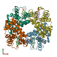
1a3n

X-ray diffraction
1.8Å resolution

DEOXY HUMAN HEMOGLOBIN

Released:
Source organism: Homo sapiens
Primary publication:
The structures of deoxy human haemoglobin and the mutant Hb Tyralpha42His at 120 K.
Acta Crystallogr D Biol Crystallogr 56 805-11 (2000)
PMID: 10930827

Function and Biology Details

Structure analysis Details

Assembly composition:
hetero tetramer (preferred)
Assembly name:
PDBe Complex ID:
PDB-CPX-159519 (preferred)
Entry contents:
2 distinct polypeptide molecules
Macromolecules (2 distinct):
Hemoglobin subunit alpha Chains: A, C
Molecule details ›
Chains: A, C
Length: 141 amino acids
Theoretical weight: 15.15 KDa
Source organism: Homo sapiens
UniProt:
  • Canonical: P69905 (Residues: 2-142; Coverage: 99%)
Gene names: HBA1, HBA2
Sequence domains: Globin
Structure domains: Globins
Hemoglobin subunit beta Chains: B, D
Molecule details ›
Chains: B, D
Length: 146 amino acids
Theoretical weight: 15.89 KDa
Source organism: Homo sapiens
UniProt:
  • Canonical: P68871 (Residues: 2-147; Coverage: 99%)
Gene name: HBB
Sequence domains: Globin
Structure domains: Globins

Ligands and Environments

1 bound ligand:
No modified residues

Experiments and Validation Details

Entry percentile scores
Spacegroup: P21
Unit cell:
a: 62.65Å b: 82.43Å c: 53.53Å
α: 90° β: 99.61° γ: 90°
R-values:
R R work R free
0.171 0.171 0.22