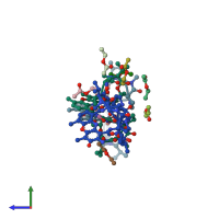
1a7y

X-ray diffraction
0.94Å resolution

CRYSTAL STRUCTURE OF ACTINOMYCIN D

Released:
Source organism: Streptomyces antibioticus
Primary publication:
Crystal Structures of Actinomycin D and Actinomycin Z3.
Angew Chem Int Ed Engl 37 2381-2384 (1998)
PMID: 29710967

Function and Biology Details

Biochemical function:
  • not assigned
Biological process:
  • not assigned
Cellular component:
  • not assigned

Structure analysis Details

Assembly composition:
monomeric (preferred)
Assembly name:
PDBe Complex ID:
PDB-CPX-162096 (preferred)
Entry contents:
1 distinct polypeptide molecule
Macromolecule:
ACTINOMYCIN D Chains: A, B, C
Molecule details ›
Chains: A, B, C
Length: 11 amino acids
Theoretical weight: 1.26 KDa
Source organism: Streptomyces antibioticus

Ligands and Environments

2 bound ligands:
3 modified residues:

Experiments and Validation Details

Entry percentile scores
X-ray source: SIEMENS
Spacegroup: P1
Unit cell:
a: 15.737Å b: 15.887Å c: 25.156Å
α: 85.93° β: 86.19° γ: 69.86°
R-values:
R R work R free
0.057 not available 0.068