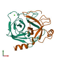
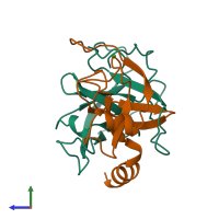
1aks

X-ray diffraction
1.8Å resolution

CRYSTAL STRUCTURE OF THE FIRST ACTIVE AUTOLYSATE FORM OF THE PORCINE ALPHA TRYPSIN

Released:
Source organism: Sus scrofa
Primary publication:
The first structure at 1.8 A resolution of an active autolysate form of porcine alpha-trysoin.
Acta Crystallogr D Biol Crystallogr 53 311-5 (1997)
PMID: 15299934

Function and Biology Details

Structure analysis Details

Assembly composition:
hetero dimer (preferred)
Assembly name:
PDBe Complex ID:
PDB-CPX-133376 (preferred)
Entry contents:
2 distinct polypeptide molecules
Macromolecules (2 distinct):
Trypsin Chain: A
Molecule details ›
Chain: A
Length: 125 amino acids
Theoretical weight: 13.3 KDa
Source organism: Sus scrofa
UniProt:
  • Canonical: P00761 (Residues: 9-133; Coverage: 54%)
Sequence domains: Trypsin
Structure domains: Trypsin-like serine proteases
Trypsin Chain: B
Molecule details ›
Chain: B
Length: 98 amino acids
Theoretical weight: 10.21 KDa
Source organism: Sus scrofa
UniProt:
  • Canonical: P00761 (Residues: 134-231; Coverage: 42%)
Sequence domains: Trypsin
Structure domains: Trypsin-like serine proteases

Ligands and Environments

1 bound ligand:
No modified residues

Experiments and Validation Details

Entry percentile scores
Spacegroup: P212121
Unit cell:
a: 77.7Å b: 53.82Å c: 47.08Å
α: 90° β: 90° γ: 90°
R-values:
R R work R free
0.2 0.2 not available