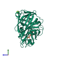
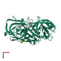
1apu

X-ray diffraction
1.8Å resolution

Crystallographic analysis of a pepstatin analogue binding to the aspartyl proteinase penicillopepsin at 1.8 angstroms resolution

Released:

Function and Biology Details

Reaction catalysed:
Hydrolysis of proteins with broad specificity similar to that of pepsin A, preferring hydrophobic residues at P1 and P1', but also cleaving 20-Gly-|-Glu-21 in the B chain of insulin. Clots milk, and activates trypsinogen.
Biological process:
Cellular component:

Structure analysis Details

Assemblies composition:
hetero dimer (preferred)
hetero tetramer
PDBe Complex ID:
PDB-CPX-133573 (preferred)
Entry contents:
2 distinct polypeptide molecules
Macromolecules (2 distinct):
Penicillopepsin-1 Chain: E
Molecule details ›
Chain: E
Length: 323 amino acids
Theoretical weight: 33.47 KDa
Source organism: Penicillium janthinellum
Expression system: Not provided
UniProt:
  • Canonical: P00798 (Residues: 1-323; Coverage: 100%)
Sequence domains: Eukaryotic aspartyl protease
Structure domains: Acid Proteases
PEPSTATIN ANALOGUE ISOVALERYL-VAL-VAL-STA-O-ET Chain: I
Molecule details ›
Chain: I
Length: 4 amino acids
Theoretical weight: 486 Da
Source organism: Penicillium janthinellum
Expression system: Not provided

Ligands and Environments

1 bound ligand:
No modified residues

Experiments and Validation Details

Entry percentile scores
Spacegroup: C2
Unit cell:
a: 97.24Å b: 46.5Å c: 65.65Å
α: 90° β: 115.34° γ: 90°
R-values:
R R work R free
0.131 not available not available
Expression system: Not provided