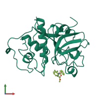
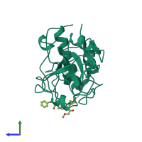
1bqi

X-ray diffraction
2.5Å resolution

USE OF PAPAIN AS A MODEL FOR THE STRUCTURE-BASED DESIGN OF CATHEPSIN K INHIBITORS. CRYSTAL STRUCTURES OF TWO PAPAIN INHIBITOR COMPLEXES DEMONSTRATE BINDING TO S'-SUBSITES.

Released:

Function and Biology Details

Reaction catalysed:
Hydrolysis of proteins with broad specificity for peptide bonds, but preference for an amino acid bearing a large hydrophobic side chain at the P2 position. Does not accept Val in P1'.
Biochemical function:
Biological process:
Cellular component:
  • not assigned

Structure analysis Details

Assembly composition:
monomeric (preferred)
Assembly name:
PDBe Complex ID:
PDB-CPX-133539 (preferred)
Entry contents:
1 distinct polypeptide molecule
Macromolecule:
Papain Chain: A
Molecule details ›
Chain: A
Length: 212 amino acids
Theoretical weight: 23.45 KDa
Source organism: Carica papaya
UniProt:
  • Canonical: P00784 (Residues: 134-345; Coverage: 65%)
Sequence domains: Papain family cysteine protease
Structure domains: Cysteine proteinases

Ligands and Environments

1 bound ligand:
No modified residues

Experiments and Validation Details

Entry percentile scores
X-ray source: SIEMENS
Spacegroup: C2
Unit cell:
a: 101Å b: 50.99Å c: 62.48Å
α: 90° β: 99.6° γ: 90°
R-values:
R R work R free
0.217 0.217 0.306