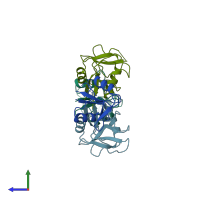
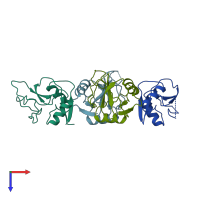
1bv4

X-ray diffraction
1.85Å resolution

APO-MANNOSE-BINDING PROTEIN-C

Released:
Source organism: Rattus norvegicus
Primary publication:
Ca2+-dependent structural changes in C-type mannose-binding proteins.
Biochemistry 37 17965-76 (1998)
PMID: 9922165

Function and Biology Details

Biochemical function:
  • not assigned
Biological process:
  • not assigned
Cellular component:
  • not assigned

Structure analysis Details

Assembly composition:
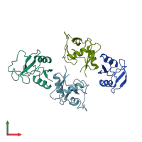
homo tetramer (preferred)
PDBe Complex ID:
PDB-CPX-140265 (preferred)
Entry contents:
1 distinct polypeptide molecule
Macromolecule:
Mannose-binding protein C Chains: A, B, C, D
Molecule details ›
Chains: A, B, C, D
Length: 118 amino acids
Theoretical weight: 13.16 KDa
Source organism: Rattus norvegicus
Expression system: Escherichia coli
UniProt:
  • Canonical: P08661 (Residues: 127-244; Coverage: 52%)
Gene name: Mbl2
Sequence domains: Lectin C-type domain
Structure domains: Mannose-Binding Protein A, subunit A

Ligands and Environments

No bound ligands
No modified residues

Experiments and Validation Details

Entry percentile scores
X-ray source: SSRL BEAMLINE BL7-1
Spacegroup: P21
Unit cell:
a: 32.39Å b: 132.62Å c: 46.81Å
α: 90° β: 94.28° γ: 90°
R-values:
R R work R free
0.212 0.212 0.256
Expression system: Escherichia coli