Function and Biology Details
Reactions catalysed:
Nucleoside triphosphate + RNA(n) = diphosphate + RNA(n+1)
Hydrolysis of four peptide bonds in the viral precursor polyprotein, commonly with Asp or Glu in the P6 position, Cys or Thr in P1 and Ser or Ala in P1'.
NTP + H(2)O = NDP + phosphate
ATP + H(2)O = ADP + phosphate
Biochemical function:
Biological process:
Cellular component:
- not assigned
Sequence domains:
Structure domains:
Structure analysis Details
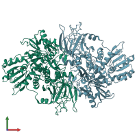
Assembly composition:
homo dimer (preferred)
Assembly name:
Serine protease/helicase NS3 (preferred)
PDBe Complex ID:
PDB-CPX-150815 (preferred)
Entry contents:
1 distinct polypeptide molecule
Macromolecule:


