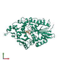
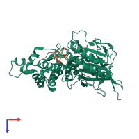
1dkp

X-ray diffraction
2.28Å resolution

CRYSTAL STRUCTURE OF PHYTATE COMPLEX OF ESCHERICHIA COLI PHYTASE AT PH 6.6. PHYTATE IS BOUND WITH ITS 3-PHOSPHATE IN THE ACTIVE SITE. HG2+ CATION ACTS AS AN INTERMOLECULAR BRIDGE

Released:
Source organism: Escherichia coli
Primary publication:
Crystal structures of Escherichia coli phytase and its complex with phytate.
Nat Struct Biol 7 108-13 (2000)
PMID: 10655611

Function and Biology Details

Structure analysis Details

Assembly composition:
monomeric (preferred)
Assembly name:
PDBe Complex ID:
PDB-CPX-139374 (preferred)
Entry contents:
1 distinct polypeptide molecule
Macromolecule:
Phytase AppA Chain: A
Molecule details ›
Chain: A
Length: 410 amino acids
Theoretical weight: 44.7 KDa
Source organism: Escherichia coli
Expression system: Escherichia coli
UniProt:
  • Canonical: P07102 (Residues: 23-432; Coverage: 100%)
Gene names: JW0963, appA, b0980
Sequence domains: Histidine phosphatase superfamily (branch 2)
Structure domains: Phosphoglycerate mutase-like

Ligands and Environments

2 bound ligands:
No modified residues

Experiments and Validation Details

Entry percentile scores
X-ray source: RIGAKU RU200
Spacegroup: P212121
Unit cell:
a: 70.573Å b: 75.127Å c: 90.393Å
α: 90° β: 90° γ: 90°
R-values:
R R work R free
0.189 0.189 0.227
Expression system: Escherichia coli