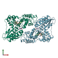
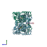
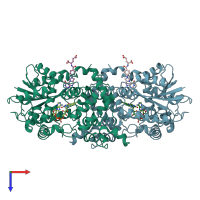
Function and Biology Details
Reaction catalysed:
Cyclobutadipyrimidine (in DNA) = 2 pyrimidine residues (in DNA)
Biochemical function:
Biological process:
Cellular component:
- not assigned
Sequence domains:
- Cryptochrome/DNA photolyase class 1
- Cryptochrome/DNA photolyase class 1, conserved site, C-terminal
- DNA photolyase, N-terminal
- Cryptochrome/DNA photolyase, FAD-binding domain
- Cryptochrome/DNA photolyase, FAD-binding domain-like superfamily
- Cryptochrome/photolyase, N-terminal domain superfamily
- Rossmann-like alpha/beta/alpha sandwich fold
Structure analysis Details
Assembly composition:
monomeric (preferred)
Assembly name:
Deoxyribodipyrimidine photo-lyase (preferred)
PDBe Complex ID:
PDB-CPX-133679 (preferred)
Entry contents:
1 distinct polypeptide molecule
Macromolecule:


