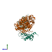
1e9y

X-ray diffraction
3Å resolution

Crystal structure of Helicobacter pylori urease in complex with acetohydroxamic acid

Released:
Source organism: Helicobacter pylori
Primary publication:
Supramolecular assembly and acid resistance of Helicobacter pylori urease.
Nat Struct Biol 8 505-9 (2001)
PMID: 11373617

Function and Biology Details

Structure analysis Details

Assembly composition:
hetero 24-mer (preferred)
PDBe Complex ID:
PDB-CPX-147152 (preferred)
Entry contents:
2 distinct polypeptide molecules
Macromolecules (2 distinct):
Urease subunit alpha Chain: A
Molecule details ›
Chain: A
Length: 238 amino acids
Theoretical weight: 26.57 KDa
Source organism: Helicobacter pylori
Expression system: Escherichia coli
UniProt:
  • Canonical: P14916 (Residues: 1-238; Coverage: 100%)
Gene names: HP_0073, hpuA, ureA
Sequence domains:
Structure domains:
Urease subunit beta Chain: B
Molecule details ›
Chain: B
Length: 569 amino acids
Theoretical weight: 61.76 KDa
Source organism: Helicobacter pylori
Expression system: Escherichia coli
UniProt:
  • Canonical: P69996 (Residues: 1-569; Coverage: 100%)
Gene names: HP_0072, hpuB, ureB
Sequence domains:
Structure domains:

Ligands and Environments

2 bound ligands:
1 modified residue:

Experiments and Validation Details

Entry percentile scores
X-ray source: PHOTON FACTORY BEAMLINE BL-6A
Spacegroup: I23
Unit cell:
a: 178.16Å b: 178.16Å c: 178.16Å
α: 90° β: 90° γ: 90°
R-values:
R R work R free
0.213 0.213 0.282
Expression system: Escherichia coli