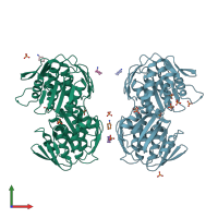
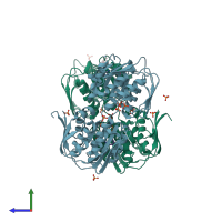
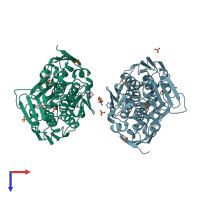
1ejd

X-ray diffraction
1.55Å resolution

Crystal structure of unliganded mura (type1)

Released:
Source organism: Enterobacter cloacae

Function and Biology Details

Reaction catalysed:
Phosphoenolpyruvate + UDP-N-acetyl-alpha-D-glucosamine = phosphate + UDP-N-acetyl-3-O-(1-carboxyvinyl)-alpha-D-glucosamine

Structure analysis Details

Assemblies composition:
monomeric (preferred)
homo tetramer
homo dimer
PDBe Complex ID:
PDB-CPX-152534 (preferred)
Entry contents:
1 distinct polypeptide molecule
Macromolecule:
UDP-N-acetylglucosamine 1-carboxyvinyltransferase Chains: A, B
Molecule details ›
Chains: A, B
Length: 419 amino acids
Theoretical weight: 44.83 KDa
Source organism: Enterobacter cloacae
Expression system: Escherichia coli
UniProt:
  • Canonical: P33038 (Residues: 1-419; Coverage: 100%)
Gene names: ECL_04571, murA, murZ
Sequence domains: EPSP synthase (3-phosphoshikimate 1-carboxyvinyltransferase)
Structure domains: Enolpyruvate transferase domain

Ligands and Environments

2 bound ligands:
1 modified residue:

Experiments and Validation Details

Entry percentile scores
X-ray source: ESRF BEAMLINE BM1A
Spacegroup: C2
Unit cell:
a: 87.177Å b: 156.297Å c: 84.036Å
α: 90° β: 91.63° γ: 90°
R-values:
R R work R free
0.185 0.185 0.207
Expression system: Escherichia coli