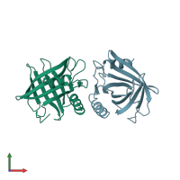
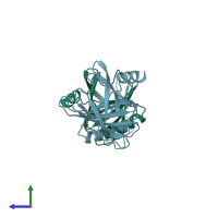
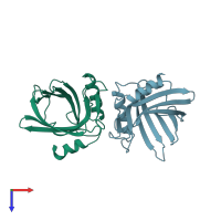
1epa

X-ray diffraction
2.1Å resolution

STRUCTURE OF THE EPIDIDYMAL RETINOIC ACID-BINDING PROTEIN AT 2.1 ANGSTROMS RESOLUTION

Released:
Source organism: Rattus norvegicus

Function and Biology Details

Biochemical function:
Biological process:
  • not assigned
Cellular component:

Structure analysis Details

Assembly composition:
homo dimer (preferred)
PDBe Complex ID:
PDB-CPX-139302 (preferred)
Entry contents:
1 distinct polypeptide molecule
Macromolecule:
Epididymal-specific lipocalin-5 Chains: A, B
Molecule details ›
Chains: A, B
Length: 164 amino acids
Theoretical weight: 18.25 KDa
Source organism: Rattus norvegicus
Expression system: Not provided
UniProt:
  • Canonical: P06911 (Residues: 23-186; Coverage: 97%)
Gene names: Erabp, Lcn5
Sequence domains: Lipocalin / cytosolic fatty-acid binding protein family
Structure domains: Lipocalin

Ligands and Environments

No bound ligands
No modified residues

Experiments and Validation Details

Entry percentile scores
Spacegroup: P21
Unit cell:
a: 39.33Å b: 58.67Å c: 66.27Å
α: 90° β: 109.34° γ: 90°
R-values:
R R work R free
0.196 0.196 not available
Expression system: Not provided