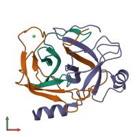
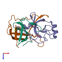
1ept

X-ray diffraction
1.8Å resolution

REFINED 1.8 ANGSTROMS RESOLUTION CRYSTAL STRUCTURE OF PORCINE EPSILON-TRYPSIN

Released:
Source organism: Sus scrofa
Primary publication:
Refined 1.8 A resolution crystal structure of the porcine epsilon-trypsin.
Biochim Biophys Acta 1209 77-82 (1994)
PMID: 7947985

Function and Biology Details

Structure analysis Details

Assembly composition:
hetero trimer (preferred)
Assembly name:
PDBe Complex ID:
PDB-CPX-133377 (preferred)
Entry contents:
3 distinct polypeptide molecules
Macromolecules (3 distinct):
Trypsin Chain: A
Molecule details ›
Chain: A
Length: 43 amino acids
Theoretical weight: 4.49 KDa
Source organism: Sus scrofa
Expression system: Not provided
UniProt:
  • Canonical: P00761 (Residues: 9-51; Coverage: 19%)
Structure domains: Trypsin-like serine proteases
Trypsin Chain: B
Molecule details ›
Chain: B
Length: 82 amino acids
Theoretical weight: 8.83 KDa
Source organism: Sus scrofa
Expression system: Not provided
UniProt:
  • Canonical: P00761 (Residues: 52-133; Coverage: 36%)
Sequence domains: Trypsin
Structure domains: Trypsin-like serine proteases
Trypsin Chain: C
Molecule details ›
Chain: C
Length: 98 amino acids
Theoretical weight: 10.21 KDa
Source organism: Sus scrofa
Expression system: Not provided
UniProt:
  • Canonical: P00761 (Residues: 134-231; Coverage: 42%)
Sequence domains: Trypsin
Structure domains: Trypsin-like serine proteases

Ligands and Environments

1 bound ligand:
No modified residues

Experiments and Validation Details

Entry percentile scores
Spacegroup: P212121
Unit cell:
a: 76.9Å b: 53.4Å c: 46.6Å
α: 90° β: 90° γ: 90°
R-values:
R R work R free
0.184 0.184 not available
Expression system: Not provided