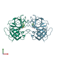
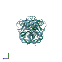
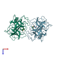
1eq9

X-ray diffraction
1.7Å resolution

CRYSTAL STRUCTURE OF FIRE ANT CHYMOTRYPSIN COMPLEXED TO PMSF

Released:
Source organism: Solenopsis invicta
Primary publication:
The structure of an insect chymotrypsin.
J. Mol. Biol. 298 895-901 (2000)
PMID: 10801356

Function and Biology Details

Structure analysis Details

Assembly composition:
homo dimer (preferred)
Assembly name:
PDBe Complex ID:
PDB-CPX-181978 (preferred)
Entry contents:
1 distinct polypeptide molecule
Macromolecule:
Chymotrypsin-1 Chains: A, B
Molecule details ›
Chains: A, B
Length: 222 amino acids
Theoretical weight: 24.04 KDa
Source organism: Solenopsis invicta
UniProt:
  • Canonical: Q7SIG2 (Residues: 1-222; Coverage: 100%)
Sequence domains: Trypsin
Structure domains: Trypsin-like serine proteases

Ligands and Environments

1 bound ligand:
No modified residues

Experiments and Validation Details

Entry percentile scores
X-ray source: RIGAKU RU200
Spacegroup: P212121
Unit cell:
a: 61.12Å b: 85.84Å c: 97.61Å
α: 90° β: 90° γ: 90°
R-values:
R R work R free
0.209 0.209 0.242