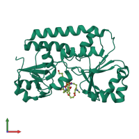
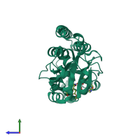
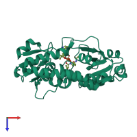
1esz

X-ray diffraction
2Å resolution

STRUCTURE OF THE PERIPLASMIC FERRIC SIDEROPHORE BINDING PROTEIN FHUD COMPLEXED WITH COPROGEN

Released:

Function and Biology Details

Structure analysis Details

Assembly composition:
monomeric (preferred)
PDBe Complex ID:
PDB-CPX-139777 (preferred)
Entry contents:
1 distinct polypeptide molecule
Macromolecule:
Iron(3+)-hydroxamate-binding protein FhuD Chain: A
Molecule details ›
Chain: A
Length: 266 amino acids
Theoretical weight: 29.69 KDa
Source organism: Escherichia coli
Expression system: Escherichia coli
UniProt:
  • Canonical: P07822 (Residues: 31-296; Coverage: 100%)
Gene names: JW0148, b0152, fhuD
Sequence domains: Periplasmic binding protein
Structure domains: Nitrogenase molybdenum iron protein domain

Ligands and Environments

1 bound ligand:
No modified residues

Experiments and Validation Details

Entry percentile scores
X-ray source: NSLS BEAMLINE X12C
Spacegroup: P63
Unit cell:
a: 86.09Å b: 86.09Å c: 91.94Å
α: 90° β: 90° γ: 120°
R-values:
R R work R free
0.219 0.219 0.241
Expression system: Escherichia coli