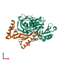
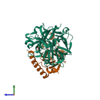
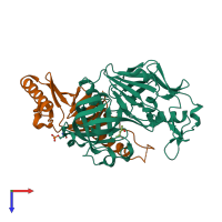
1f34

X-ray diffraction
2.45Å resolution

CRYSTAL STRUCTURE OF ASCARIS PEPSIN INHIBITOR-3 BOUND TO PORCINE PEPSIN

Released:

Function and Biology Details

Reaction catalysed:
Preferential cleavage: hydrophobic, preferably aromatic, residues in P1 and P1' positions. Cleaves 1-Phe-|-Val-2, 4-Gln-|-His-5, 13-Glu-|-Ala-14, 14-Ala-|-Leu-15, 15-Leu-|-Tyr-16, 16-Tyr-|-Leu-17, 23-Gly-|-Phe-24, 24-Phe-|-Phe-25 and 25-Phe-|-Tyr-26 bonds in the B chain of insulin.
Biological process:
Cellular component:

Structure analysis Details

Assembly composition:
hetero dimer (preferred)
PDBe Complex ID:
PDB-CPX-133554 (preferred)
Entry contents:
2 distinct polypeptide molecules
Macromolecules (2 distinct):
Pepsin A Chain: A
Molecule details ›
Chain: A
Length: 326 amino acids
Theoretical weight: 34.59 KDa
Source organism: Sus scrofa
UniProt:
  • Canonical: P00791 (Residues: 60-385; Coverage: 88%)
Gene name: PGA
Sequence domains: Eukaryotic aspartyl protease
Structure domains: Acid Proteases
Major pepsin inhibitor 3 Chain: B
Molecule details ›
Chain: B
Length: 149 amino acids
Theoretical weight: 16.41 KDa
Source organism: Ascaris suum
Expression system: Escherichia coli
UniProt:
  • Canonical: P19400 (Residues: 21-169; Coverage: 100%)
Sequence domains: Pepsin inhibitor-3-like repeated domain
Structure domains: Pepsin inhibitor-3

Ligands and Environments

1 bound ligand:
1 modified residue:

Experiments and Validation Details

Entry percentile scores
X-ray source: RIGAKU RU200
Spacegroup: C2221
Unit cell:
a: 65.374Å b: 97.593Å c: 136.394Å
α: 90° β: 90° γ: 90°
R-values:
R R work R free
0.218 0.218 0.29
Expression system: Escherichia coli