Function and Biology Details
Reaction catalysed:
Random hydrolysis of (1->4)-linkages between N-acetyl-beta-D-glucosamine and D-glucuronate residues in hyaluronate
Biochemical function:
Biological process:
Cellular component:
Sequence domains:
Structure domain:
Structure analysis Details
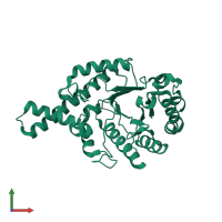
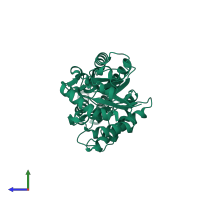
Assembly composition:
monomeric (preferred)
Assembly name:
Hyaluronidase (preferred)
PDBe Complex ID:
PDB-CPX-170598 (preferred)
Entry contents:
1 distinct polypeptide molecule
Macromolecule:
Ligands and Environments
No bound ligands
No modified residues
Experiments and Validation Details
X-ray source:
EMBL/DESY, HAMBURG BEAMLINE BW7A
Spacegroup:
P21
Expression system: Trichoplusia ni


