


Function and Biology Details
Biochemical function:
Biological process:
Cellular component:
Sequence domains:
- ATP synthase, F1 complex, gamma subunit
- ATP synthase, F1 complex, delta/epsilon subunit
- F0F1 ATP synthase delta/epsilon subunit, N-terminal
- ATP synthase, F1 complex, gamma subunit superfamily
- ATP synthase epsilon subunit, C-terminal domain
- ATP synthase delta/epsilon subunit, C-terminal domain superfamily
- ATP synthase, F1 complex, delta/epsilon subunit, N-terminal
Structure analysis Details
Assemblies composition:
Assembly name:
ATP synthase (preferred)
PDBe Complex ID:
PDB-CPX-141305 (preferred)
Entry contents:
2 distinct polypeptide molecules
Macromolecules (2 distinct):
ATP synthase epsilon chain
Molecule details ›
Chain: E
Length: 138 amino acids
Theoretical weight: 14.96 KDa
Source organism: Escherichia coli
UniProt:
Sequence domains:
Length: 138 amino acids
Theoretical weight: 14.96 KDa
Source organism: Escherichia coli
UniProt:
- Canonical:
 P0A6E6 (Residues: 2-139; Coverage: 99%)
Sequence domains:
- ATP synthase, Delta/Epsilon chain, beta-sandwich domain
- ATP synthase, Delta/Epsilon chain, long alpha-helix domain
ATP synthase gamma chain
Molecule details ›
Chain: G
Length: 230 amino acids
Theoretical weight: 25.68 KDa
Source organism: Escherichia coli
UniProt:
Sequence domains: ATP synthase
Structure domains:
Length: 230 amino acids
Theoretical weight: 25.68 KDa
Source organism: Escherichia coli
UniProt:
- Canonical: P0ABA6 (Residues: 19-248; Coverage: 80%)
Sequence domains: ATP synthase
Structure domains:
Ligands and Environments
No bound ligands
No modified residues
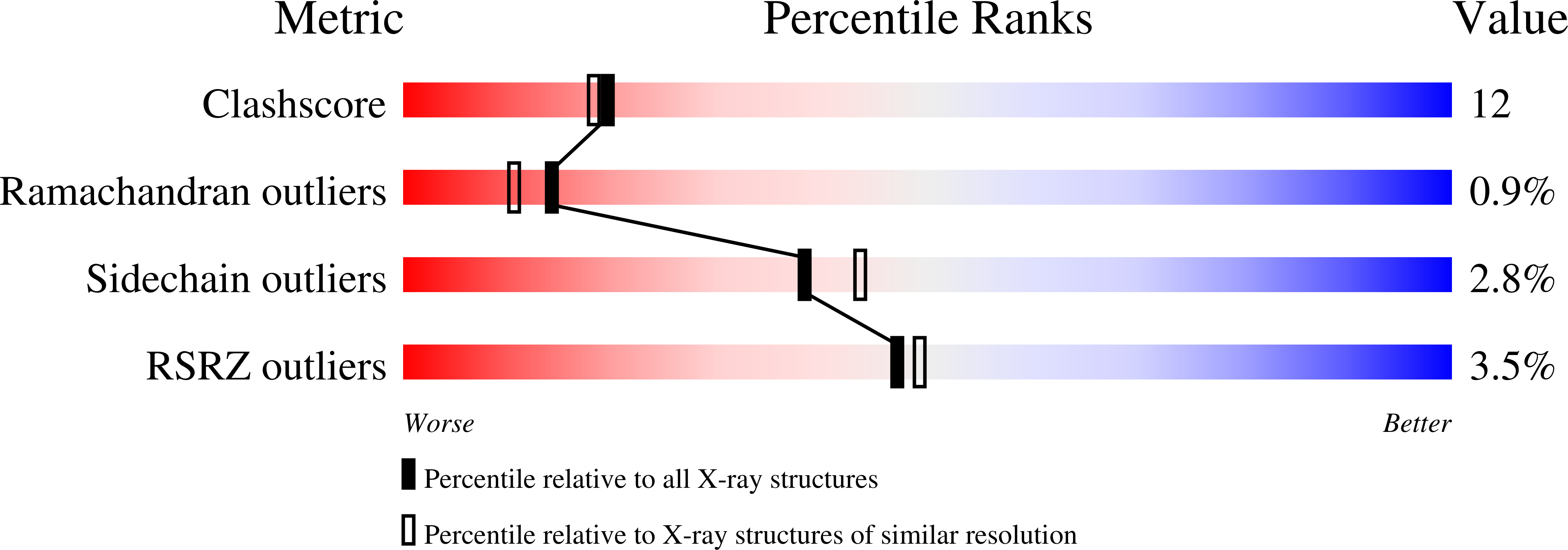
Experiments and Validation Details
wwPDB Validation report is not available for this entry.
X-ray source:
APS BEAMLINE 14-BM-C
Spacegroup:
C2221
