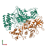
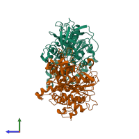
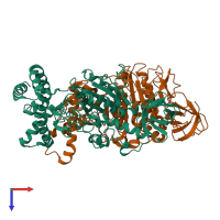
Function and Biology Details
Reaction catalysed:
ATP + H(2)O + 4 H(+)(Side 1) = ADP + phosphate + 4 H(+)(Side 2)
Biochemical function:
Biological process:
Cellular component:
Sequence domains:
- P-loop containing nucleoside triphosphate hydrolase
- ATP synthase, F1 complex, alpha subunit
- ATP synthase, F1 complex, beta subunit
- ATP synthase, alpha subunit, C-terminal
- ATPase, alpha/beta subunit, nucleotide-binding domain, active site
- ATPase, F1/V1/A1 complex, alpha/beta subunit, nucleotide-binding domain
- ATPase, F1/V1/A1 complex, alpha/beta subunit, N-terminal domain
- ATPase, F1/V1/A1 complex, alpha/beta subunit, N-terminal domain superfamily
5 more domains
Structure analysis Details
Assembly composition:
hetero hexamer (preferred)
Assembly name:
ATP synthase chloroplastic (preferred)
PDBe Complex ID:
PDB-CPX-133608 (preferred)
Entry contents:
2 distinct polypeptide molecules
Macromolecules (2 distinct):
Ligands and Environments
No bound ligands
No modified residues
Experiments and Validation Details
X-ray source:
EMBL/DESY, HAMBURG BEAMLINE X11
Spacegroup:
R32


